
Recent Advances in 64Cu/67Cu-Based Radiopharmaceuticals
 , ,
, ,
Abstract
:1. Introduction
2. Summary of Copper-Based Radiopharmaceuticals, Reported in 2018–2023
3. Copper-Based Radiopharmaceuticals Based on Peptides
3.1. Octreotate
3.2. PSMA
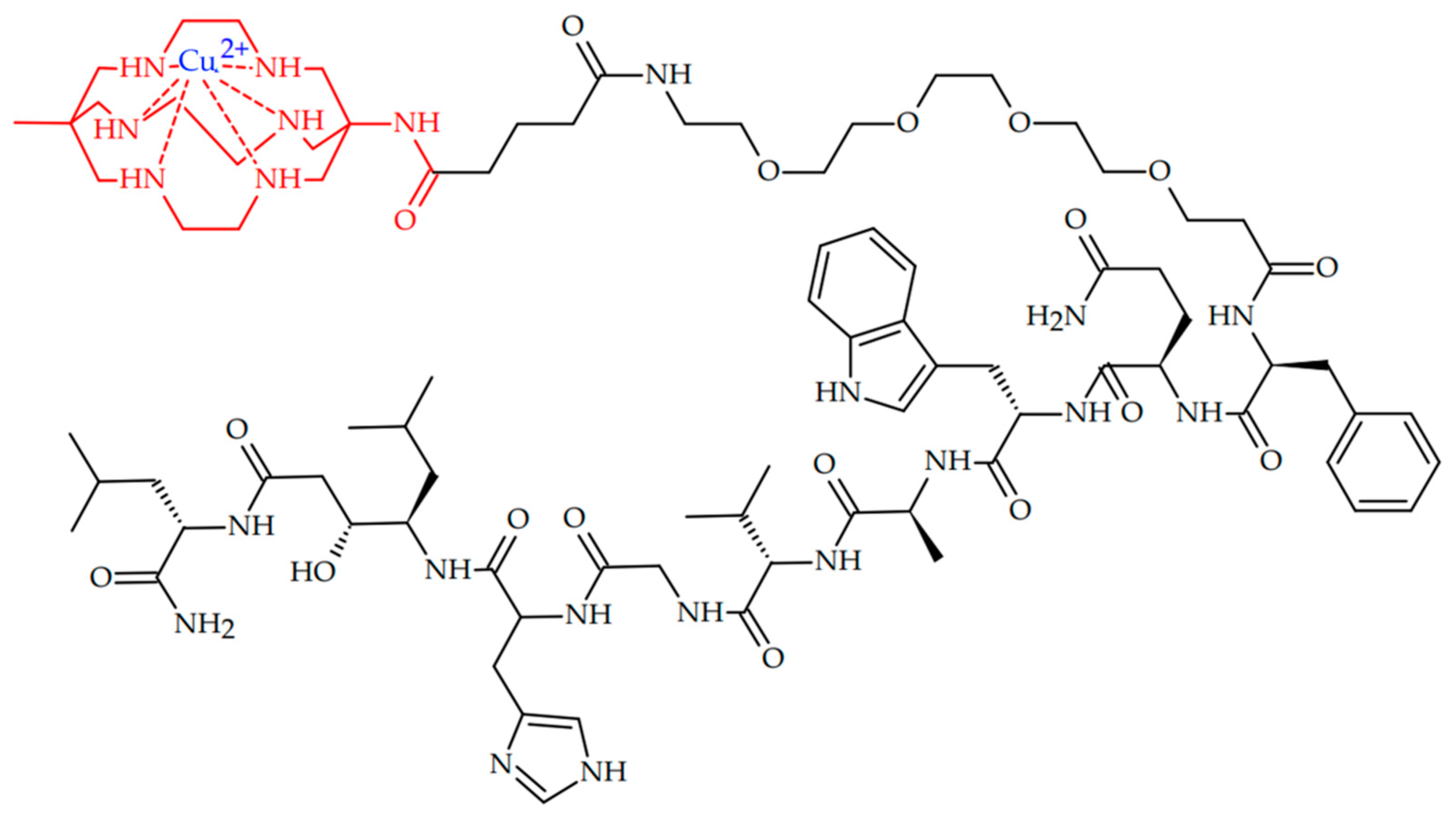
3.3. Other Peptides
4. Copper-Based Radiopharmaceuticals for Radioimmunotherapy
4.1. Direct Conjugation of Radiolabeled Chelator and Antibody
4.2. Pretargeting Approach in Conjugation of Radiolabeled Chelator and Antibody
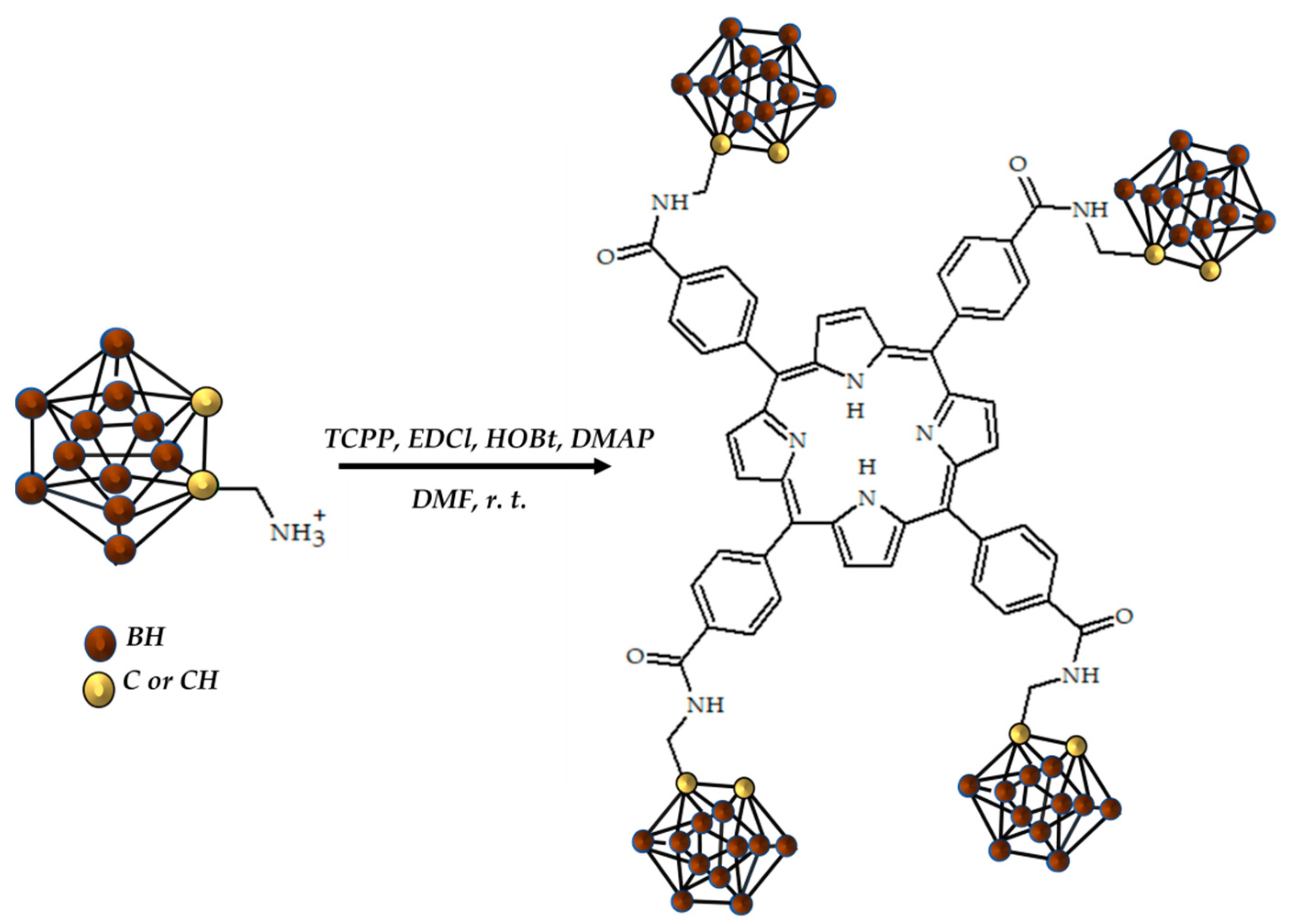
5. Another Copper-Based Radiopharmaceutical
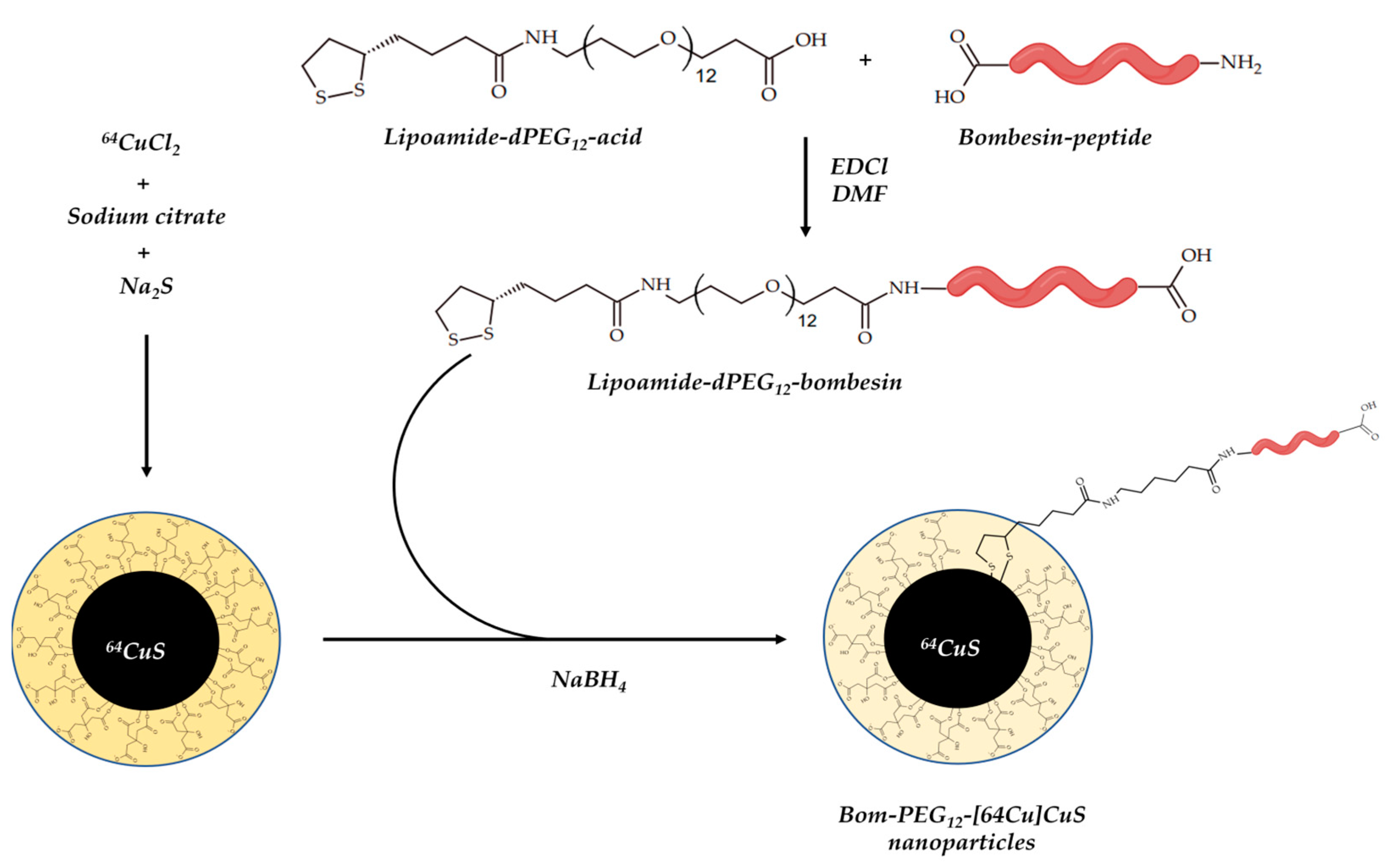
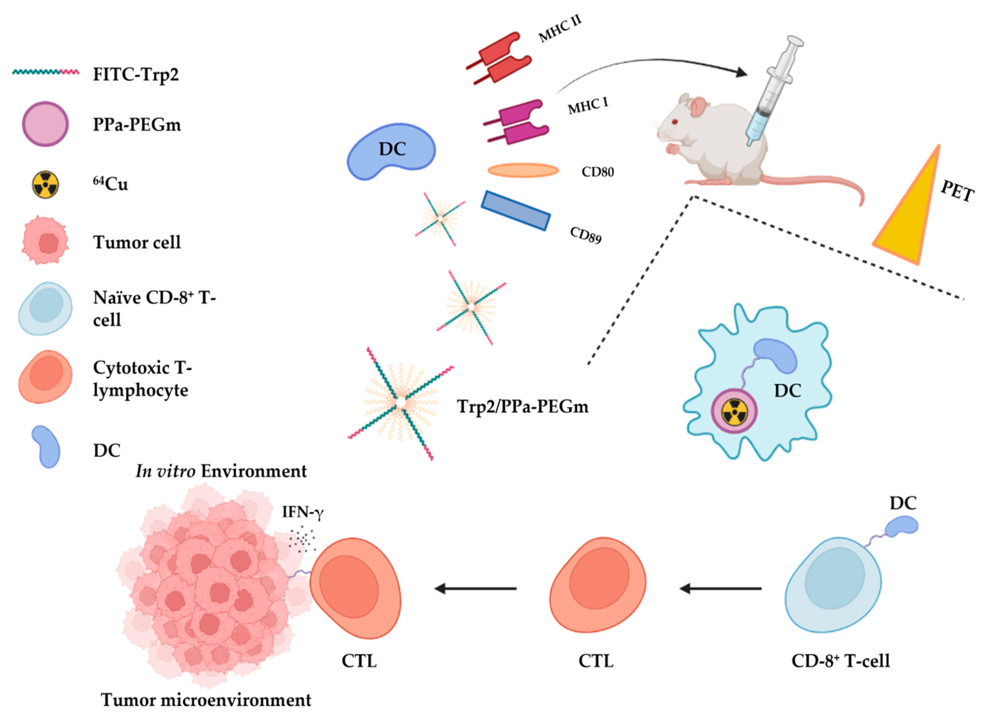
Nanoparticles (Nps)
6. Conclusions
Funding
Conflicts of Interest
References
- Baskar, R.; Dai, J.; Wenlong, N.; Yeo, R.; Yeoh, K.W. Biological response of cancer cells to radiation treatment. Front. Mol. Biosci. 2014, 1, 24. [Google Scholar] [CrossRef] [PubMed] [Green Version]
- Nickoloff, J.; Sharma, N.; Taylor, L. Biological consequences of radiation-induced DNA damage: Relevance to radiotherapy. Genes 2013, 11, 99. [Google Scholar] [CrossRef] [PubMed] [Green Version]
- Crișan, G.; Moldovean-Cioroianu, N.S.; Timaru, D.G.; Andrieș, G.; Căinap, C.; Chiș, V. Radiopharmaceuticals for PET and SPECT imaging: A literature review over the last decade. Int. J. Mol. Sci. 2022, 23, 5023. [Google Scholar] [CrossRef] [PubMed]
- Yeong, C.H.; Cheng, M.H.; Ng, K.H. Therapeutic radionuclides in nuclear medicine: Current and future prospects. J. Zhejiang Univ. 2014, 15, 845–863. [Google Scholar] [CrossRef] [Green Version]
- Boschi, A.; Martini, P.; Costa, V.; Pagnoni, A.; Uccelli, L. Interdisciplinary tasks in the cyclotron production of Radiometals for medical applications. The case of 47Sc as example. Molecules 2019, 24, 444. [Google Scholar] [CrossRef] [Green Version]
- Currie, G.M.; Wheat, J.M.; Davidson, R.; Kiat, H. Radionuclide production. Radiographer 2011, 58, 46–52. [Google Scholar] [CrossRef]
- Onda, Y.; Taniguchi, K.; Yoshimura, K.; Kato, H.; Takahashi, J.; Wakiyama, Y.; Coppin, F.; Smith, H. Radionuclides from the Fukushima Daiichi nuclear power plant in terrestrial systems. Nat. Rev. Earth Environ. 2020, 1, 644–660. [Google Scholar] [CrossRef]
- Zhang, T.; Das, S.K.; Fels, D.R.; Hansen, K.S.; Wong, T.Z.; Dewhirst, M.W.; Vlahovic, G. PET with 62Cu-ATSM and 62Cu-PTSM is a useful imaging tool for hypoxia and perfusion in pulmonary lesions. Am. J. Roentgenol. 2013, 201, W698. [Google Scholar] [CrossRef] [Green Version]
- Richardson, M.P.; Koepp, M.J.; Brooks, D.J.; Duncan, J.S. 11C-flumazenil PET in neocortical epilepsy. Neurology 1998, 51, 485–492. [Google Scholar] [CrossRef]
- Zürcher, N.R.; Walsh, E.C.; Phillips, R.D.; Cernasov, P.M.; Tseng, C.E.; Dharanikota, A.; Smith, E.; Li, Z.; Kinard, J.L.; Bizzell, J.C.; et al. A simultaneous [11C] raclopride positron emission tomography and functional magnetic resonance imaging investigation of striatal dopamine binding in autism. Transl. Psychiatry 2021, 11, 33. [Google Scholar] [CrossRef]
- Sharma, R.; D’Souza, M.; Jaimini, A.; Hazari, P.P.; Saw, S.; Pandey, S.; Singh, D.; Solanki, Y.; Kumar, N.; Mishra, A.K.; et al. A comparison study of 11C-methionine and 18F-fluorodeoxyglucose positron emission tomography-computed tomography scans in evaluation of patients with recurrent brain tumors. Ind. J. Nucl. Med. 2016, 31, 93. [Google Scholar] [CrossRef] [Green Version]
- US Food and Drug Administration. FDA approves 11C-choline for PET in prostate cancer. J. Nucl. Med. 2012, 53, 11N. [Google Scholar]
- Kitajima, K.; Abe, K.; Takeda, M.; Yoshikawa, H.; Ohigashi, M.; Osugi, K.; Koyama, H.; Yamakado, K. Clinical impact of 11C-Pittsburgh compound-B positron emission tomography in addition to magnetic resonance imaging and single-photon emission computed tomography on diagnosis of mild cognitive impairment to Alzheimer’s disease. Medicine 2021, 100, e23969. [Google Scholar] [CrossRef]
- Lewis, J.S.; Laforest, R.; Dehdashti, F.; Grigsby, P.W.; Welch, M.J.; Siegel, B.A. An imaging comparison of 64Cu-ATSM and 60Cu-ATSM in cancer of the uterine cervix. J. Nucl. Med. 2008, 49, 1177–1182. [Google Scholar] [CrossRef] [PubMed] [Green Version]
- Mbakaza, O.; Vangu, M.D. 18F-FDG PET/CT Imaging: Normal Variants, Pitfalls, and Artifacts Musculoskeletal, Infection, and Inflammation. Front. Nucl. Med. 2022, 2, 847810. [Google Scholar] [CrossRef]
- Tatum, J.L.; Kalen, J.D.; Jacobs, P.M.; Riffle, L.A.; James, A.; Thang, L.; Sanders, C.; Hollingshead, M.G.; Basuli, F.; Shi, J.; et al. 3′-[18F] fluoro-3′-deoxythymidine ([18F] FLT) Positron Emission Tomography as an In Vivo Biomarker of inhibition of CDK 4/6-Rb pathway by Palbociclib in a patient derived bladder tumor. J. Transl. Med. 2022, 20, 375. [Google Scholar] [CrossRef] [PubMed]
- US Food and Drug Administration. FDA Approves New 68Ga Kit for Prostate Cancer PET. J. Nucl. Med. 2022, 63, 26N. [Google Scholar]
- Mohamad, H.; Ali, S.; Emmanuel, P. The role of 68Ga-DOTA-NOC PET/CT in evaluating neuroendocrine tumors. Nucl. Med. Commun. 2017, 38, 170–177. [Google Scholar]
- Bruvoll, R.; Blakkisrud, J.; Mikalsen, L.T.; Connelly, J.; Stokke, C. Correlations between [68Ga] Ga-DOTA-TOC uptake and absorbed dose from [177Lu] Lu-DOTA-TATE. Cancers 2023, 15, 1134. [Google Scholar] [CrossRef]
- Kratochwil, C.; Flechsig, P.; Lindner, T.; Abderrahim, L.; Altmann, A.; Mier, W.; Adeberg, S.; Rathke, H.; Röhrich, M.; Winter, H.; et al. 68Ga-FAPI PET/CT: Tracer uptake in 28 different kinds of cancer. J. Nucl. Med. 2019, 60, 801–805. [Google Scholar] [CrossRef] [Green Version]
- Jalilian, A.; Rostampour, N.; Rowshanfarzad, P.; Shafaii, K.; Kamali-Dehghan, M.; Akhlaghi, M. Preclinical studies of [Cu] ATSM as a PET radiopharmaceutical for fibrosarcoma imaging. Acta Pharm. 2009, 59, 45–55. [Google Scholar] [CrossRef] [PubMed]
- Becker, K.; Schwartz, P.; Aluicio-Sarduy, E.; Jeffery, J.; Massey, C.; Hernandez, R.; Ronnekleiv-Kelly, S.; Pirasteh, A.; Engle, J. Preclinical Evaluation of 43Sc-FAPI PET for Detection of Pancreatic Ductal Adenocarcinoma. J. Nucl. Med. 2022, 63 (Suppl. S2), 2616. [Google Scholar]
- Juget, F.; Durán, T.; Nedjadi, Y.; Talip, Z.; Grundler, P.V.; Favaretto, C.; Casolaro, P.; Dellepiane, G.; Braccini, S.; Bailat, C.; et al. Activity Measurement of 44Sc and Calibration of Activity Measurement Instruments on Production Sites and Clinics. Molecules 2023, 28, 1345. [Google Scholar] [CrossRef] [PubMed]
- Hernandez, R.; Valdovinos, H.F.; Yang, Y.; Chakravarty, R.; Hong, H.; Barnhart, T.E.; Cai, W. 44Sc: An attractive isotope for peptide-based PET imaging. Mol. Pharm. 2014, 11, 2954–2961. [Google Scholar] [CrossRef] [PubMed]
- Pfeifer, A.; Knigge, U.; Binderup, T.; Mortensen, J.; Oturai, P.; Loft, A.; Berthelsen, A.K.; Langer, S.W.; Rasmussen, P.; Elema, D.; et al. 64Cu-DOTATATE PET for neuroendocrine tumors: A prospective head-to-head comparison with 111In-DTPA-octreotide in 112 patients. J. Nucl. Med. 2015, 56, 847–854. [Google Scholar] [CrossRef] [PubMed] [Green Version]
- Kaalep, A.; Huisman, M.; Sera, T.; Vugts, D.; Boellaard, R.; EARL; EATRIS; TRISTAN Consortium (# IB4SD-116106). Feasibility of PET/CT system performance harmonisation for quantitative multicentre 89Zr studies. EJNMMI Phys. 2018, 5, 26. [Google Scholar] [CrossRef] [Green Version]
- Joyce van Sluis, J.; Boellaard, R.; Dierckx, R.A.; van Esch, E.L.; Croes, D.A.; de Ruijter, L.K.; van de Donk, P.P.; de Vries, E.G.; Noordzij, W.; Brouwers, A.H. Optimisation of scan duration and image quality in oncological 89Zr immunoPET imaging using the Biograph Vision PET/CT. Eur. J. Nucl. Med. Mol. Imaging 2023, 1–3. [Google Scholar] [CrossRef]
- Yoon, J.K.; Park, B.N.; Ryu, E.K.; An, Y.S.; Lee, S.J. Current perspectives on 89Zr-PET imaging. Int. J. Mol. Sci. 2020, 21, 4309. [Google Scholar] [CrossRef]
- Zeglis, B.M.; Lewis, J.S. The Bioconjugation and Radiosynthesis of Zr-89-DFO-labeled Antibodies. JOVE-J. Vis. Exp. 2015, 96. [Google Scholar]
- Such, B.M.; Pantuck, A.J.; Bernhard, J.C.; Morris, M.A.; Master, V.A.; Scott, A.M.; Van Praet, C.; Bailly, C.; Aksoy, T.; Merkx, R.; et al. Results from phase 3 study of 89Zr-DFO-girentuximab for PET/CT imaging of clear cell renal cell carcinoma (ZIRCON). J. Clin. Oncol. 2023, 41, LBA602. [Google Scholar] [CrossRef]
- Kuker, R.; Sztejnberg, M.; Gulec, S. I-124 imaging and dosimetry. Mol. Imaging Radiat. Ther. 2017, 26 (Suppl. S1), 66. [Google Scholar] [CrossRef] [PubMed]
- Yang, F.; Yang, Z.; Feng, J.; Zhang, L.; Daqing Ma, M.D.; Jigang Yang, M.D. Three phase bone scintigraphy with Tc-MDP and serological indices in detecting infection after internal fixation in malunion or nonunion traumatic fractures. Hell. J. Nucl. Med. 2016, 19, 130–134. [Google Scholar] [PubMed]
- Fuster, D.; Maurel, J.; Muxi, A.; Setoain, X.; Ayuso, C.; Martin, F.; Ortega, M.L.; Fuertes, S.; Pons, F. Is there a role for 99mTc-anti-CEA monoclonal antibody imaging in the diagnosis of recurrent colorectal carcinoma? Q. J. Nucl. Med. 2003, 47, 109–115. [Google Scholar]
- de Graaf, W.H.; Heger, M.; van Ginhoven, T.M.; van Cappellen, G.; Bennink, R.J.; Kullak-Ublick, G.A.; Hesselmann, R.; van Gulik, T.M.; Stieger, B.J. Transporters involved in the hepatic uptake of 99mTc-mebrofenin and indocyanine green. J. Hepatol. 2011, 54, 738–745. [Google Scholar] [CrossRef] [PubMed] [Green Version]
- Park, H.M. 123I: Almost a Designer Radioiodine for Thyroid Scanning. J. Nucl. Med. 2002, 43, 77–78. [Google Scholar]
- Tolosa, E.; Borght, T.V.; Moreno, E. DaTSCAN Clinically Uncertain Parkinsonian Syndromes Study Group. Accuracy of DaTSCAN (123I-ioflupane) SPECT in diagnosis of patients with clinically uncertain parkinsonism: 2-Year follow-up of an open-label study. Mov. Disord. 2007, 22, 2346–2351. [Google Scholar] [CrossRef]
- Druckenbrod, R.W.; Williams, C.C.; Gelfand, M.J. Iofetamine hydrochloride I 123: A new radiopharmaceutical for cerebral perfusion imaging. DICP 1989, 23, 19–24. [Google Scholar] [CrossRef]
- Chopra, A. [123I]-2-Iodo-2-amino-3-phenylpropanoic acid. In Molecular Imaging and Contrast Agent Database (MICAD) [Internet]; National Center for Biotechnology Information (US): Bethesda, MD, USA, 2008; pp. 2004–2013. [Google Scholar]
- Wafelman, A.R.; Hoefnagel, C.A.; Maes, R.A.; Beijnen, J.H. Radioiodinated metaiodobenzylguanidine: A review of its biodistribution and pharmacokinetics, drug interactions, cytotoxicity and dosimetry. Eur J. Nucl. Med. 1994, 21, 545–559. [Google Scholar] [CrossRef]
- Hoffer, P. Gallium: Mechanisms. J. Nucl. Med. 1980, 21, 282–285. [Google Scholar]
- Rizvi, T.; Deng, C.; Rehm, P. Indium-111 capromab pendetide (ProstaScint®) demonstrates renal cell carcinoma and aortocaval nodal metastases from prostate adenocarcinoma. World J. Nucl. Med. 2015, 14, 209–211. [Google Scholar] [CrossRef]
- Iagaru, A.; Gambhir, S.S.; Goris, M.L. 90Y-ibritumomab therapy in refractory non-Hodgkin’s lymphoma: Observations from 111In-ibritumomab pretreatment imaging. J. Nucl. Med. 2008, 49, 1809–1812. [Google Scholar] [CrossRef] [PubMed] [Green Version]
- Maini, C.L.; Bergomi, S.; Romano, L.; Sciuto, R. 153Sm-EDTMP for bone pain palliation in skeletal metastases. Eur. J. Nucl. Med. Mol. Imaging 2004, 31, S171–S178. [Google Scholar] [CrossRef] [PubMed]
- International Atomic Energy Agency. Therapeutic Radiopharmaceuticals Labelled with Copper-67, Rhenium-186 and Scandium-47; IAEA-TECDOC-1945; IAEA: Vienna, Austria, 2021. [Google Scholar]
- Ehrhardt, G.J.; Ketring, A.R.; Cutler, C.S. Radioisotope radiotherapy research and achievements at the University of Missouri Research Reactor. Czechoslov. J. Phys. 2003, 53, A707–A712. [Google Scholar] [CrossRef]
- Siwowska, K.; Guzik, P.; Domnanich, K.A.; Monné Rodríguez, J.M.; Bernhardt, P.; Ponsard, B.; Hasler, R.; Borgna, F.; Schibli, R.; Köster, U.; et al. Therapeutic potential of 47Sc in comparison to 177Lu and 90Y: Preclinical investigations. Pharmaceutics 2019, 11, 424. [Google Scholar] [CrossRef] [Green Version]
- Phillips, W.T.; Goins, B.; Bao, A.; Vargas, D.; Guttierez, J.E.; Trevino, A.; Miller, J.R.; Henry, J.; Zuniga, R.; Vecil, G.; et al. Rhenium-186 liposomes as convection-enhanced nanoparticle brachytherapy for treatment of glioblastoma. Neuro Oncol. 2012, 14, 416–425. [Google Scholar] [CrossRef] [PubMed] [Green Version]
- Argyrou, M.; Valassi, A.; Andreou, M.; Lyra, M. Dosimetry and therapeutic ratios for rhenium-186 HEDP. Int. Sch. Res. Not. 2013, 2013, 124603. [Google Scholar] [CrossRef]
- Bocher, M.M.D.; Shibley, N.C.N.T.; Chisin, R.M.D. The Use of Xenon-133 Ventilation Scan Performed Immediately After Tc-99m MAA Perfusion Scan. Clin. Nucl. Med. 1993, 18, 157–158. [Google Scholar] [CrossRef]
- del Olmo-García, M.I.; Prado-Wohlwend, S.; Bello, P.; Segura, A.; Merino-Torres, J.F. Peptide receptor radionuclide therapy with [177Lu] Lu-DOTA-TATE in patients with advanced GEP NENS: Present and future directions. Cancers 2022, 14, 584. [Google Scholar] [CrossRef]
- Kayano, D.; Kinuya, S. Current consensus on I-131 MIBG therapy. Nucl. Med. Mol. Imaging 2018, 52, 254–265. [Google Scholar] [CrossRef]
- MedlinePlus [Internet]; National Library of Medicine (US): Bethesda, MD, USA. Available online: https://go.drugbank.com/drugs/DB09498 (accessed on 1 July 2020).
- Ahenkorah, S.; Cassells, I.; Deroose, C.M.; Cardinaels, T.; Burgoyne, A.R.; Bormans, G.; Ooms, M.; Cleeren, F. Bismuth-213 for targeted radionuclide therapy: From atom to bedside. Pharmaceutics 2021, 13, 599. [Google Scholar] [CrossRef]
- Delpassand, E.; Tworowska, I.; Shanoon, F.; Nunez, R.; Flores II, L.; Muzammil, A.; Stallons, T.; Saidi, A.; Torgue, J. First clinical experience using targeted alpha-emitter therapy with Pb-212-DOTAMTATE (AlphaMedix TM) in patients with SSTR(+) neuroendocrine tumors. J. Nucl. Med. 2019, 60, 559. [Google Scholar]
- Ma, J.; Li, L.; Liao, T.; Gong, W.; Zhang, C. Efficacy and Safety of 225Ac-PSMA-617-Targeted Alpha Therapy in Metastatic Castration-Resistant Prostate Cancer: A Systematic Review and Meta-Analysis. Front. Oncol. 2022, 12, 796657. [Google Scholar] [CrossRef] [PubMed]
- Satapathy, S.; Sood, A.; Das, C.K.; Mittal, B.R. Evolving role of 225Ac-PSMA radioligand therapy in metastatic castration-resistant prostate cancer—A systematic review and meta-analysis. Prostate Cancer Prostatic Dis. 2021, 24, 880–890. [Google Scholar] [CrossRef] [PubMed]
- Maguire, W.F.; McDevitt, M.R.; Smith-Jones, P.M.; Scheinberg, D.A. Efficient 1-step radiolabeling of monoclonal antibodies to high specific activity with 225Ac for α-particle radioimmunotherapy of cancer. J. Nucl. Med. 2014, 55, 1492–1498. [Google Scholar] [CrossRef] [Green Version]
- Karlsson, J.; Hagemann, U.B.; Schatz, C.; Grant, D.; Ellingsen, C.; Kristian, A.; Mihaylova, D.; Uran, S.R.; Suominen, M.; Bjerke, R.M.; et al. HER2-targeted thorium-227 conjugate (HER2-TTC): Efficacy in a HER2 positive orthotopic bone model. Cancer Res. 2017, 77 (Suppl. S13), 5857. [Google Scholar] [CrossRef]
- Miller, C.; Rousseau, J.; Ramogida, C.F.; Celler, A.; Rahmim, A.; Uribe, C.F. Implications of physics, chemistry and biology for dosimetry calculations using theranostic pairs. Theranostics 2022, 12, 232–259. [Google Scholar] [CrossRef]
- Qaim, S.M.; Scholten, B.; Neumaier, B. New developments in the production of theranostic pairs of radionuclides. J. Radioanal. Nucl. Chem. 2018, 318, 1493–1509. [Google Scholar] [CrossRef] [Green Version]
- Fonseca, A.I.; Alves, V.H.; do Carmo, S.J.; Silva, M.; Hrynchak, I.; Alves, F.; Falcão, A.; Abrunhosa, A.J. Production of GMP-Compliant Clinical Amounts of Copper-61 Radiopharmaceuticals from Liquid Targets. Pharmaceutics 2022, 15, 723. [Google Scholar] [CrossRef]
- Jødal, L.; Le Loirec, C.; Champion, C. Positron range in PET imaging: An alternative approach for assessing and correcting the blurring. Phys. Med. Bio 2012, 57, 3931. [Google Scholar] [CrossRef] [Green Version]
- Sanchez-Crespo, A.; Andreo, P.; Larsson, S.A. Positron flight in human tissues and its influence on PET image spatial resolution. Eur. J. Nucl. Med. Mol. Imaging 2004, 31, 44–51. [Google Scholar] [CrossRef]
- Wei, W.; Rosenkrans, Z.T.; Liu, J.; Huang, G.; Luo, Q.Y.; Cai, W. ImmunoPET: Concept, design, and applications. Chem. Rev. 2020, 120, 3787–3851. [Google Scholar] [CrossRef] [PubMed]
- IAEA Radioisotopes and Radiopharmaceuticals Series No. 7. Copper-64 Radiopharmaceuticals: Production, Quality Control and Clinical Applications. Available online: https://www.iaea.org/publications/14727/copper-64-radiopharmaceuticals-production-quality-control-and-clinical-applications (accessed on 19 May 2023).
- Thieme, S.; Walther, M.; Pietzsch, H.J.; Henniger, J.; Preusche, S.; Mäding, P.; Steinbach, J. Module-assisted preparation of 64Cu with high specific activity. Appl. Radiat. Isot. 2012, 70, 602–608. [Google Scholar] [CrossRef]
- Jauregui-Osoro, M.; De Robertis, S.; Halsted, P.; Gould, S.M.; Yu, Z.; Paul, R.L.; Marsden, P.K.; Gee, A.D.; Fenwick, A.; Blower, P.J. Production of copper-64 using a hospital cyclotron: Targetry, purification and quality analysis. Nucl. Med. Commun. 2021, 42, 1024–1038. [Google Scholar] [CrossRef] [PubMed]
- Zinn, K.R.; Chaudhuri, T.R.; Cheng, T.P.; Steven Morris, J.; Meyer, W.A., Jr. Production of no-carrier-added 64Cu from zinc metal irradiated under boron shielding. Cancer 1994, 73 (Suppl. S3), 774–778. [Google Scholar] [CrossRef]
- Chakravarty, R.; Shetty, P.; Nair, K.V.; Rajeswari, A.; Jagadeesan, K.C.; Sarma, H.D.; Rangarajan, V.; Krishnatry, R.; Chakraborty, S. Reactor produced [64Cu] CuCl2 as a PET radiopharmaceutical for cancer imaging: From radiochemistry laboratory to nuclear medicine clinic. Ann. Nucl. Med. 2020, 34, 899–910. [Google Scholar] [CrossRef]
- Dellepiane, G.; Casolaro, P.; Mateu, I.; Scampoli, P.; Braccini, S. Alternative routes for 64Cu production using an 18 MeV medical cyclotron in view of theranostic applications. Appl. Radiat. Isot. 2023, 191, 110518. [Google Scholar] [CrossRef] [PubMed]
- Delpassand, E.S.; Ranganathan, D.; Wagh, N.; Shafie, A.; Gaber, A.; Abbasi, A.; Kjaer, A.; Tworowska, I.; Núñez, R. 64Cu-DOTATATE PET/CT for imaging patients with known or suspected somatostatin receptor–positive neuroendocrine tumors: Results of the first US prospective, reader-masked clinical trial. J. Nucl. Med. 2020, 61, 890–896. [Google Scholar] [CrossRef]
- Braune, A.; Oehme, L.; Freudenberg, R.; Hofheinz, F.; van den Hoff, J.; Kotzerke, J.; Hoberück, S. Comparison of image quality and spatial resolution between 18F, 68Ga, and 64Cu phantom measurements using a digital Biograph Vision PET/CT. EJNMMI Phys. 2022, 9, 1–5. [Google Scholar]
- Srivastava, S.C. A Bridge not too Far: Personalized Medicine with the use of Theragnostic Radiopharmaceuticals. J. Postgrad. Med. Educ. Res. 2013, 47, 31–46. [Google Scholar] [CrossRef]
- Ali, S.K.; Khandaker, M.U.; Al-Mugren, K.S.; Latif, S.A.; Bradley, D.A.; Okhunov, A.A.; Sulieman, A. Evaluation of production cross-sections for theranostic 67Cu radionuclide via proton-induced nuclear reaction on 68Zn target. Appl. Radiat. Isot. 2021, 173, 109735. [Google Scholar] [CrossRef]
- Mou, L.; Martini, P.; Pupillo, G.; Cieszykowska, I.; Cutler, C.S.; Mikołajczak, R. 67Cu production capabilities: A mini review. Molecules 2022, 27, 1501. [Google Scholar] [CrossRef] [PubMed]
- Brühlmann, S.A.; Walther, M.; Kreller, M.; Reissig, F.; Pietzsch, H.J.; Kniess, T.; Kopka, K. Cyclotron-Based Production of 67Cu for Radionuclide Theranostics via the 70Zn (p, α) 67Cu Reaction. Pharmaceutics 2023, 16, 314. [Google Scholar] [CrossRef]
- Kozempel, J.; Abbas, K.; Simonelli, F.; Bulgheroni, A.; Holzwarth, U.; Gibson, N. Preparation of 67Cu via deuteron irradiation of 70Zn. Radioch Acta 2012, 100, 419–424. [Google Scholar] [CrossRef]
- Nigron, E.; Guertin, A.; Haddad, F.; Sounalet, T. Is 70Zn (d, x) 67Cu the best way to produce 67Cu for medical applications? Front. Med. 2021, 8, 674617. [Google Scholar] [CrossRef]
- O’Brien, H.A., Jr. The preparation of 67Cu from 67Zn in a nuclear reactor. Int. J. App. Radiat. Isot. 1969, 20, 121–124. [Google Scholar] [CrossRef]
- Merrick, M.J.; Rotsch, D.A.; Tiwari, A.; Nolen, J.; Brossard, T.; Song, J.; Wadas, T.J.; Sunderland, J.J.; Graves, S.A. Imaging and dosimetric characteristics of 67Cu. Phys. Med. Bio 2021, 66, 035002. [Google Scholar] [CrossRef]
- Merrick, M.J.; Rotsch, D.A.; Tiwari, A.; Nolen, J.; Brossard, T.; Song, J.; Wadas, T.J.; Sunderland, J.J.; Graves, S.A. Half-life of 67Cu. J. Phys. Commun. 2021, 5, 085007. [Google Scholar] [CrossRef]
- Zhou, Y.; Li, J.; Xu, X.; Zhao, M.; Zhang, B.; Deng, S.; Wu, Y. 64Cu-based radiopharmaceuticals in molecular imaging. Technol. Cancer Res. Treat. 2019, 18, 1533033819830758. [Google Scholar] [CrossRef] [PubMed] [Green Version]
- Miranda, V.M. Medicinal inorganic chemistry: An updated review on the status of metallodrugs and prominent metallodrug candidates. Rev. Inorg. Chem. 2022, 42, 29–52. [Google Scholar] [CrossRef]
- Liu, T.; Karlsen, M.; Karlberg, A.M.; Redalen, K.R. Hypoxia imaging and theranostic potential of [64Cu][Cu (ATSM)] and ionic Cu (II) salts: A review of current evidence and discussion of the retention mechanisms. EJNMMI Res. 2020, 10, 33. [Google Scholar] [CrossRef] [Green Version]
- Krasnovskaya, O.; Spector, D.; Zlobin, A.; Pavlov, K.; Gorelkin, P.; Erofeev, A.; Beloglazkina, E.; Majouga, A. Metals in Imaging of Alzheimer’s Disease. Int. J. Mol. Sci. 2020, 21, 9190. [Google Scholar] [CrossRef] [PubMed]
- Ahmedova, A.; Todorov, B.; Burdzhiev, N.; Goze, C. Copper radiopharmaceuticals for theranostic applications. Eur. J. Med. Chem. 2018, 157, 1406–1425. [Google Scholar] [CrossRef] [PubMed]
- Capriotti, G.; Piccardo, A.; Giovannelli, E.; Signore, A. Targeting Copper in Cancer Imaging and Therapy: A New Theragnostic Agent. J. Clin. Med. 2023, 12, 223. [Google Scholar] [CrossRef] [PubMed]
- Johnbeck, C.B.; Knigge, U.; Loft, A.; Berthelsen, A.K.; Mortensen, J.; Oturai, P.; Langer, S.W.; Elema, D.R.; Kjaer, A. Head-to-head comparison of 64Cu-DOTATATE and 68Ga-DOTATOC PET/CT: A prospective study of 59 patients with neuroendocrine tumors. J. Nucl. Med. 2017, 58, 451–457. [Google Scholar] [CrossRef] [Green Version]
- Cullinane, C.; Jeffery, C.M.; Roselt, P.D.; Dam, E.M.; Jackson, S.; Kuan, K.; Jackson, P.; Binns, D.; van Zuylekom, J.; Harris, M.J.; et al. Peptide receptor radionuclide therapy with 67Cu-CuSarTATE is highly efficacious against a somatostatin-positive neuroendocrine tumor model. J. Nucl. Med. 2020, 61, 1800–1805. [Google Scholar] [CrossRef]
- Song, H.; Guja, K.E.; Yang, E.J.; Guo, H.H. 64Cu-DOTATATE Uptake in a Pulmonary Hamartoma. Clin. Nucl. Med. 2022, 10, 1097. [Google Scholar] [CrossRef]
- Umbricht, C.A.; Benešová, M.; Hasler, R.; Schibli, R.; van der Meulen, N.P.; Müller, C. Design and preclinical evaluation of an albumin-binding PSMA ligand for 64Cu-based PET imaging. Mol. Pharm. 2018, 15, 5556–5564. [Google Scholar] [CrossRef]
- Kelly, J.M.; Ponnala, S.; Amor-Coarasa, A.; Zia, N.A.; Nikolopoulou, A.; Williams, C., Jr.; Schlyer, D.J.; DiMagno, S.G.; Donnelly, P.S.; Babich, J.W. Preclinical evaluation of a high-affinity sarcophagine-containing PSMA ligand for 64Cu/67Cu-based theranostics in prostate cancer. Mol. Pharm. 2020, 17, 1954–1962. [Google Scholar] [CrossRef]
- Zia, N.; Cullinane, C.; Zuylekom, K.; Waldeck, K.; McInnes, E.; Buncic, G.; Haskali, M.; Roselt, P.; Hicks, R.; Donnelly, P. A Bivalent Inhibitor of Prostate Specific Membrane Antigen Radiolabeled with Copper-64 with High Tumor Uptake and Retention. Angew. Chem. Int. Ed. 2019, 58, 14991–14994. [Google Scholar] [CrossRef]
- McInnes, L.E.; Cullinane, C.; Roselt, P.D.; Jackson, S.; Blyth, B.J.; van Dam, E.M.; Zia, N.A.; Harris, M.J.; Hicks, R.J.; Donnelly, P.S. Therapeutic efficacy of a bivalent inhibitor of prostate-specific membrane antigen labeled with 67Cu. J. Nucl. Med. 2021, 62, 829–832. [Google Scholar] [CrossRef]
- Sarkar, S.; Bhatt, N.; Ha, Y.S.; Huynh, P.T.; Soni, N.; Lee, W.; Lee, Y.J.; Kim, J.Y.; Pandya, D.N.; An, G.I.; et al. High in vivo stability of 64Cu-labeled cross-bridged chelators is a crucial factor in improved tumor imaging of RGD peptide conjugates. J. Med. Chem. 2018, 61, 385–395. [Google Scholar] [CrossRef] [PubMed]
- Qiao, Z.; Xu, J.; Gonzalez, R.; Miao, Y. Novel 64Cu-labeled NOTA-conjugated lactam-cyclized alpha-melanocyte-stimulating hormone peptides with enhanced tumor to kidney uptake ratios. Mol. Pharm. 2022, 19, 2535–2541. [Google Scholar] [CrossRef] [PubMed]
- Huynh, T.T.; van Dam, E.M.; Sreekumar, S.; Mpoy, C.; Blyth, B.J.; Muntz, F.; Harris, M.J.; Rogers, B.E. Copper-67-Labeled Bombesin Peptide for Targeted Radionuclide Therapy of Prostate Cancer. Pharmaceutics 2022, 15, 728. [Google Scholar] [CrossRef] [PubMed]
- Xu, M.; Han, Y.; Liu, G.; Xu, Y.; Duan, D.; Liu, H.; Du, F.; Luo, P.; Liu, Z. Preclinical study of a fully human anti-PD-L1 antibody as a theranostic agent for cancer immunotherapy. Mol. Pharm. 2018, 15, 4426–4433. [Google Scholar] [CrossRef]
- Lee, W.; Sarkar, S.; Pal, R.; Kim, J.Y.; Park, H.; Huynh, P.T.; Bhise, A.; Bobba, K.N.; Kim, K.I.; Ha, Y.S.; et al. Successful Application of CuAAC Click Reaction in Constructing 64Cu-Labeled Antibody Conjugates for Immuno-PET Imaging. ACS Appl. Bio Mater. 2021, 4, 2544–2557. [Google Scholar] [CrossRef] [PubMed]
- Hao, G.; Mastren, T.; Silvers, W.; Hassan, G.; Öz, O.; Sun, X. Copper-67 radioimmunotheranostics for simultaneous immunotherapy and immuno-SPECT. Sci. Rep. 2021, 11, 3622. [Google Scholar] [CrossRef] [PubMed]
- Clausen, A.S.; Christensen, C.; Christensen, E.; Cold, S.; Kristensen, L.K.; Hansen, A.E.; Kjaer, A. Development of a 64Cu-labeled CD4+ T cell targeting PET tracer: Evaluation of CD4 specificity and its potential use in collagen-induced arthritis. EJNMMI Res. 2022, 12, 62. [Google Scholar] [CrossRef]
- Keinänen, O.; Fung, K.; Brennan, J.M.; Zia, N.; Harris, M.; van Dam, E.; Biggin, C.; Hedt, A.; Stoner, J.; Donnelly, P.S.; et al. Harnessing 64Cu/67Cu for a theranostic approach to pretargeted radioimmunotherapy. Proc. Natl. Acad. Sci. USA 2020, 117, 28316–28327. [Google Scholar] [CrossRef]
- Jallinoja, V.I.; Carney, B.D.; Bhatt, K.; Abbriano, C.H.; Schlyer, D.J.; Yazaki, P.J.; Houghton, J.L. Investigation of Copper-64-Based Host–Guest Chemistry Pretargeted Positron Emission Tomography. Mol. Pharm. 2022, 19, 2268–2278. [Google Scholar] [CrossRef]
- Shi, Y.; Li, J.; Zhang, Z.; Duan, D.; Zhang, Z.; Liu, H.; Liu, T.; Liu, Z. Tracing boron with fluorescence and positron emission tomography imaging of boronated porphyrin nanocomplex for imaging-guided boron neutron capture therapy. ACS Appl. Mater. Interfaces 2018, 10, 43387–43395. [Google Scholar] [CrossRef]
- Earley, D.F.; Flores, J.E.; Guillou, A.; Holland, J.P. Photoactivatable bis (thiosemicarbazone) derivatives for copper-64 radiotracer synthesis. Dalton Trans. 2022, 51, 5041–5052. [Google Scholar] [CrossRef] [PubMed]
- Cai, H.; Xie, F.; Mulgaonkar, A.; Chen, L.; Sun, X.; Hsieh, J.T.; Peng, F.; Tian, R.; Li, L.; Wu, C.; et al. Bombesin functionalized 64Cu-copper sulfide nanoparticles for targeted imaging of orthotopic prostate cancer. Nanomedicine 2018, 13, 1695–1705. [Google Scholar] [CrossRef] [PubMed]
- Madru, R.; Budassi, M.; Benveniste, H.; Lee, H.; Smith, S.D.; Schlyer, D.J.; Vaska, P.; Knutsson, L.; Strand, S.E. Simultaneous preclinical positron emission tomography-magnetic resonance imaging study of lymphatic drainage of chelator-free 64Cu-labeled nanoparticles. Cancer Biother. Radiopharm. 2018, 33, 213–220. [Google Scholar] [CrossRef] [PubMed]
- Lee, H.; Gaddy, D.; Ventura, M.; Bernards, N.; de Souza, R.; Kirpotin, D.; Wickham, T.; Fitzgerald, J.; Zheng, J.; Hendriks, B.S. Companion diagnostic 64Cu-liposome positron emission tomography enables characterization of drug delivery to tumors and predicts response to cancer. Theranostics 2018, 8, 2300. [Google Scholar] [CrossRef]
- Thakare, V.; Tran, V.L.; Natuzzi, M.; Thomas, E.; Moreau, M.; Romieu, A.; Collin, B.; Courteau, A.; Vrigneaud, J.M.; Louis, C.; et al. Functionalization of theranostic AGuIX® nanoparticles for PET/MRI/optical imaging. RSC Adv. 2019, 9, 24811–24815. [Google Scholar] [CrossRef] [Green Version]
- Zhou, H.; Zhang, Q.; Cheng, Y.; Xiang, L.; Shen, G.; Wu, X.; Cai, H.; Li, D.; Zhu, H.; Zhang, R.; et al. 64Cu-labeled melanin nanoparticles for PET/CT and radionuclide therapy of tumor. Nanomed. Nanotechnol. Biol. Med. 2020, 29, 102248. [Google Scholar] [CrossRef]
- Paiva, I.; Mattingly, S.; Wuest, M.; Leier, S.; Vakili, M.R.; Weinfeld, M.; Lavasanifar, A.; Wuest, F. Synthesis and analysis of 64Cu-labeled GE11-modified polymeric micellar nanoparticles for EGFR-targeted molecular imaging in a colorectal cancer model. Mol. Pharm. 2020, 17, 1470–1481. [Google Scholar] [CrossRef]
- He, Z.; Jia, H.; Zheng, M.; Wang, H.; Yang, W.; Gao, L.; Zhang, Z.; Xue, J.; Xu, B.; Yang, W.; et al. Trp2 peptide-assembled nanoparticles with intrinsically self-chelating 64Cu properties for PET imaging tracking and dendritic cell-based immunotherapy against melanoma. ACS Appl. Bio Mater. 2021, 4, 5707–5716. [Google Scholar] [CrossRef]
- Rogoza, O.; Megnis, K.; Kudrjavceva, M.; Gerina-Berzina, A.; Rovite, V. Role of Somatostatin Signalling in Neuroendocrine Tumours. Int. J. Mol. Sci. 2022, 23, 1447. [Google Scholar] [CrossRef]
- Sanli, Y.; Garg, I.; Kandathil, A.; Kendi, T.; Zanetti, M.J.; Kuyumcu, S.; Subramaniam, R.M. Neuroendocrine tumor diagnosis and management: 68Ga-DOTATATE PET/CT. Am. J. Roentgenol. 2018, 211, 267–277. [Google Scholar] [CrossRef] [Green Version]
- Urso, L.; Nieri, A.; Uccelli, L.; Castello, A.; Artioli, P.; Cittanti, C.; Marzola, M.C.; Florimonte, L.; Castellani, M.; Bissoli, S.; et al. Lutathera® Orphans: State of the Art and Future Application of Radioligand Therapy with 177Lu-DOTATATE. Pharmaceutics 2023, 15, 1110. [Google Scholar] [CrossRef] [PubMed]
- Hennrich, U.; Benešová, M. [68Ga] Ga-DOTA-TOC: The first FDA-approved 68Ga-radiopharmaceutical for PET imaging. Pharmaceutics 2020, 13, 38. [Google Scholar] [CrossRef] [PubMed] [Green Version]
- Vahidfar, N.; Farzanehfar, S.; Abbasi, M.; Mirzaei, S.; Delpassand, E.S.; Abbaspour, F.; Salehi, Y.; Biersack, H.J.; Ahmadzadehfar, H. Diagnostic Value of Radiolabelled Somatostatin Analogues for Neuroendocrine Tumour Diagnosis: The Benefits and Drawbacks of [64Cu] Cu-DOTA-TOC. Cancers 2022, 14, 1914. [Google Scholar] [CrossRef]
- Machulkin, A.E.; Shafikov, R.R.; Uspenskaya, A.A.; Petrov, S.A.; Ber, A.P.; Skvortsov, D.A.; Nimenko, E.A.; Zyk, N.U.; Smirnova, G.B.; Pokrovsky, V.S.; et al. Synthesis and biological evaluation of PSMA ligands with aromatic residues and fluorescent conjugates based on them. J. Med. Chem. 2021, 64, 4532–4552. [Google Scholar] [CrossRef] [PubMed]
- Machulkin, A.E.; Ivanenkov, Y.A.; Aladinskaya, A.V.; Veselov, M.S.; Aladinskiy, V.A.; Beloglazkina, E.K.; Koteliansky, V.E.; Shakhbazyan, A.G.; Sandulenko, Y.B.; Majouga, A.G. Small-molecule PSMA ligands. Current state, SAR and perspectives. J. Drug Target. 2016, 24, 679–693. [Google Scholar] [CrossRef] [PubMed]
- Debnath, S.; Zhou, N.; McLaughlin, M.; Rice, S.; Pillai, A.K.; Hao, G.; Sun, X. PSMA-targeting imaging and theranostic agents—Current status and future perspective. Int. J. Mol. Sci. 2022, 23, 1158. [Google Scholar] [CrossRef]
- Jeitner, T.M.; Babich, J.W.; Kelly, J.M. Advances in PSMA theranostics. Trans. Oncol. 2022, 22, 101450. [Google Scholar] [CrossRef]
- U.S. Food and Drug Administration. Drug Approval Package: Illuccix; U.S. Food and Drug Administration (FDA): Washington, DC, USA, 2022.
- Locametz EPAR. European Medicines Agency (EMA). 12 October 2022. Available online: https://www.ema.europa.eu/en/medicines/human/EPAR/locametz (accessed on 19 May 2023).
- Milot, M.C.; Benesty, O.B.; Dumulon-Perreault, V.; Ait-Mohand, S.; Richard, P.O.; Rousseau, É.; Guérin, B. 64Cu-DOTHA2-PSMA, a Novel PSMA PET Radiotracer for Prostate Cancer with a Long Imaging Time Window. Pharmaceutics 2022, 15, 996. [Google Scholar] [CrossRef]
- Kelly, J.; Amor-Coarasa, A.; Ponnala, S.; Nikolopoulou, A.; Williams, C.; Schlyer, D.; Zhao, Y.; Kim, D.; Babich, J.W. Trifunctional PSMA-targeting constructs for prostate cancer with unprecedented localization to LNCaP tumors. Eur. J. Nucl. Med. Mol. Imaging 2018, 45, 1841–1851. [Google Scholar] [CrossRef]
- Yang, J.; Xu, J.; Gonzalez, R.; Lindner, T.; Kratochwil, C.; Miao, Y. 68Ga-DOTA-GGNle-CycMSHhex targets the melanocortin-1 receptor for melanoma imaging. Sci. Trans. Med. 2018, 10, eaau4445. [Google Scholar] [CrossRef] [Green Version]
- Rurarz, B.P.; Bukowczyk, M.; Gibka, N.; Piastowska-Ciesielska, A.W.; Karczmarczyk, U.; Ulański, P. Nanostrategies for Therapeutic and Diagnostic Targeting of Gastrin-Releasing Peptide Receptor. Int. J. Mol. Sci. 2023, 24, 3455. [Google Scholar] [CrossRef]
- Nayak, T.K.; Brechbiel, M.W. Radioimmunoimaging with longer-lived positron-emitting radionuclides: Potentials and challenges. Bioconjugate Chem. 2009, 20, 825–841. [Google Scholar] [CrossRef] [PubMed] [Green Version]
- Larson, S.M.; Carrasquillo, J.A.; Cheung, N.K.V.; Press, O.W. Radioimmunotherapy of human tumours. Nat. Rev. Cancer 2015, 15, 347–360. [Google Scholar] [CrossRef] [PubMed] [Green Version]
- Bailly, C.; Cléry, P.F.; Faivre-Chauvet, A.; Bourgeois, M.; Guérard, F.; Haddad, F.; Bodet-Milin, C. Immuno-PET for Clinical Theranostic Approaches. Int. J. Mol. Sci. 2016, 18, 57. [Google Scholar] [CrossRef] [PubMed] [Green Version]
- Liu, J.; Chen, Z.; Li, Y.; Zhao, W.; Wu, J.; Zhang, Z. PD-1/PD-L1 checkpoint inhibitors in tumor immunotherapy. Front. Pharm. 2021, 12, 731798. [Google Scholar] [CrossRef]
- De Sousa Linhares, A.; Battin, C.; Jutz, S.; Leitner, J.; Hafner, C.; Tobias, J.; Wiedermann, U.; Kundi, M.; Zlabinger, G.J.; Grabmeier-Pfistershammer, K.; et al. Therapeutic PD-L1 antibodies are more effective than PD-1 antibodies in blocking PD-1/PD-L1 signaling. Sci. Rep. 2019, 9, 11472. [Google Scholar] [CrossRef] [Green Version]
- Vu, T.; Claret, F.X. Trastuzumab: Updated mechanisms of action and resistance in breast cancer. Front. Oncol. 2012, 2, 62. [Google Scholar] [CrossRef] [Green Version]
- Capelan, M.; Pugliano, L.; De Azambuja, E.; Bozovic, I.; Saini, K.S.; Sotiriou, C.; Piccart-Gebhart, M.J. Pertuzumab: New hope for patients with HER2-positive breast cancer. Ann. Oncol. 2012, 24, 273–282. [Google Scholar] [CrossRef]
- Yap, H.Y.; Tee, S.; Wong, M.; Chow, S.K.; Peh, S.C.; Teow, S.Y. Pathogenic Role of Immune Cells in Rheumatoid Arthritis: Implications in Clinical Treatment and Biomarker Development. Cells 2018, 7, 161. [Google Scholar] [CrossRef] [Green Version]
- Van de Watering, F.C.J.; Rijpkema, M.; Robillard, M.; Oyen, W.J.G.; Boerman, O.C. Pretargeted Imaging and Radioimmunotherapy of Cancer Using Antibodies and Bioorthogonal Chemistry. Front. Med. 2014, 1, 44. [Google Scholar] [CrossRef] [Green Version]
- Jeon, W.S.; Moon, K.; Park, S.H.; Chun, H.; Ko, Y.H.; Lee, J.Y.; Kim, K. Complexation of Ferrocene Derivatives by the Cucurbit[7]uril Host: A Comparative Study of the Cucurbituril and Cyclodextrin Host Families. J. Am. Chem. Soc. 2005, 127, 12984–12989. [Google Scholar] [CrossRef] [PubMed]
- Mohajershojai, T.; Jha, P.; Boström, A.; Frejd, F.Y.; Yazaki, P.J.; Nestor, M. In Vitro Characterization of 177Lu-DOTA-M5A Anti-Carcinoembryonic Antigen Humanized Antibody and HSP90 Inhibition for Potentiated Radioimmunotherapy of Colorectal Cancer. Front. Oncol. 2022, 12, 849338. [Google Scholar] [CrossRef]
- Malouff, T.D.; Seneviratne, D.S.; Ebner, D.K.; Stross, W.C.; Waddle, M.R.; Trifiletti, D.M.; Krishnan, S. Boron neutron capture therapy: A review of clinical applications. Front. Oncol. 2021, 11, 601820. [Google Scholar] [CrossRef]
- Fukumitsu, N.; Matsumoto, Y. Development of an Imaging Technique for Boron Neutron Capture Therapy. Cells 2021, 10, 2135. [Google Scholar] [CrossRef]
- Moran, M.; MacDonald, T.; Liu, T.; Forbes, J.; Zheng, G.; Valliant, J. Copper-64-labeled porphysomes for PET imaging. J. Nucl. Med. 2014, 55, 1016. [Google Scholar]
- Wani, S.U.; Ali, M.; Masoodi, M.H.; Khan, N.A.; Zargar, M.I.; Hassan, R.; Mir, S.A.; Gautam, S.P.; Gangadharappa, H.V.; Osmani, R.M. A review on nanoparticles categorization, characterization and applications in drug delivery systems. Vib. Spectrosc. 2022, 121, 103407. [Google Scholar] [CrossRef]
- Baig, N.; Kammakakam, I.; Falath, W. Nanomaterials: A review of synthesis methods, properties, recent progress, and challenges. Mater. Adv. 2021, 2, 1821–1871. [Google Scholar] [CrossRef]
- Shreyash, N.; Bajpai, S.; Khan, M.A.; Vijay, Y.; Tiwary, S.K.; Sonker, M. Green synthesis of nanoparticles and their biomedical applications: A review. ACS Appl. Nano Mater. 2021, 4, 11428–11457. [Google Scholar] [CrossRef]
- Mohanraj, V.J.; Chen, Y.J. Nanoparticles—A review. Trop. J. Pharm. Res. 2006, 5, 561–573. [Google Scholar] [CrossRef] [Green Version]
- Forte, E.; Fiorenza, D.; Torino, E.; Costagliola di Polidoro, A.; Cavaliere, C.; Netti, P.A.; Salvatore, M.; Aiello, M. Radiolabeled PET/MRI nanoparticles for tumor imaging. J. Clin. Med. 2019, 9, 89. [Google Scholar] [CrossRef] [Green Version]
- Pijeira, M.S.; Viltres, H.; Kozempel, J.; Sakmár, M.; Vlk, M.; İlem-Özdemir, D.; Ekinci, M.; Srinivasan, S.; Rajabzadeh, A.R.; Ricci-Junior, E.; et al. Radiolabeled nanomaterials for biomedical applications: Radiopharmacy in the era of nanotechnology. EJNMMI Radiopharm. Chem. 2022, 7, 8. [Google Scholar] [CrossRef]
- Carrese, B.; Sanità, G.; Lamberti, A. Nanoparticles Design for Theranostic Approach in Cancer Disease. Cancers 2022, 14, 4654. [Google Scholar] [CrossRef] [PubMed]
- Zhou, M.; Zhang, R.; Huang, M.; Lu, W.; Song, S.; Melancon, M.P.; Tian, M.; Liang, D.; Li, C. A chelator-free multifunctional [64Cu] CuS nanoparticle platform for simultaneous micro-PET/CT imaging and photothermal ablation therapy. J. Am. Chem. Soc. 2010, 132, 15351–15358. [Google Scholar] [CrossRef] [PubMed] [Green Version]
- Lux, F.; Mignot, A.; Mowat, P.; Louis, C.; Dufort, S.; Bernhard, C.; Denat, F.; Boschetti, F.; Brunet, C.; Antoine, R.; et al. Ultrasmall rigid particles as multimodal probes for medical applications. Angew. Chem. Int. Ed. 2011, 50, 12299–12303. [Google Scholar] [CrossRef] [PubMed]
- Fan, Q.; Cheng, K.; Hu, X.; Ma, X.; Zhang, R.; Yang, M.; Lu, X.; Xing, L.; Huang, W.; Gambhir, S.S.; et al. Transferring biomarker into molecular probe: Melanin nanoparticle as a naturally active platform for multimodality imaging. J. Am. Chem. Soc. 2014, 136, 15185–15194. [Google Scholar] [CrossRef] [Green Version]


| Positron-Emitting Radionuclides | ||||||
|---|---|---|---|---|---|---|
| Nuclide | Decay | Energy, keV (Intensity) | Production | Drugs | Target/Application | Ref. |
| Copper-62 (62Cu) | T1/2 = 9.76 min β+ = 97.8% EC = 2.2% | β+ mean energy 1319: 2937 (max, 97.6%), 1321 (av); γ: 511 (195.6%); | 62Zn/62Cu generator Cyclotron 61Ni(p,γ)62Cu | [62Cu]ATSM [62Cu]PTSM | Tumor hypoxia | [8] |
| Carbon-11 (11C) | T1/2 = 20.4 min β+ = 99.8% | β+ mean energy 385.7: 960 (max, 99.8%), 385.7 (av); γ: 511 (195.5%); | Cyclotron 14N(p,α)11C | [11C]Flumazenil | GABA, epilepsy imaging | [9] |
| [11C]Raclopride | D2 dopamine receptors, Parkinson Disease imaging | [10] | ||||
| [11C]Methionine | Neuroncology, tumor imaging | [11] | ||||
| [11C]Choline | Prostate cancer | [12] | ||||
| [11C]Pittsburgh Compound B | Alzheimer disease | [13] | ||||
| Copper-60 (60Cu) | T1/2 = 23.7 min β+ = 93% EC = 7% | β+ mean energy 970: 1912 (max, 11.6%), 839.6 (av); 1982 (max, 49.0%), 872.0 (av); 2947 (max, 15.0%), 1104 (av); γ: 511 (185.0%), 826.4 (21.7%), 1332.5 (88.0%), 1791.6 (45.4%); | Cyclotron 60Ni(p,n)60Cu | [60Cu]ATSM | Tumor hypoxia | [14] |
| Fluorine-18 (18F) | T1/2 = 109.8 min β+ = 96.7% EC = 3.3% | β+ mean energy 249.8: 633.5 (max, 96.7%), 249.8 (av); γ: 511 (193.5%); | Cyclotron 18O(p,n) 18F | [18F] FDG (18F-2-fluoro-2-deoxy-D-glucose) | Tumors Neurological disorders Inflammation | [15] |
| [18F]FLT (18F-3′-fluoro-3′-deoxythymidine | Thymidine kinase-1 (TK-1) imaging biomarker of cellular proliferation Cancer | [16] | ||||
| Gallium-68 (68Ga) | T1/2 = 67.7 min β+ = 88.9% EC = 11.1% | β+ mean energy 829.5: 1899 (max, 87.7%), 836.0 (av); γ: 511 (177.8%), 1077 (3.22%) | 68Zn(p,n) 68Ga 68Ge/68Ga generator | [68Ga]PSMA-11; [68Ga]DOTA-TATE; [68Ga]DOTA-TOC; [68Ga]DOTA-NOC; [68Ga]FAPI-04 | Prostate cancer imaging SSTR(+) neuroendocrine tumors imaging Fibroblast-activation-protein—positive tumor imaging | [17,18,19,20] |
| Copper-61 (61Cu) | T1/2 = 3.34 h β+ = 61% EC = 39% | β+ mean energy 500: 932.8 (max, 5.8%), 399.0 (av); 1216 (max, 51.0%), 524.0 (av); γ: 511 (123%), 283.0 (12.7%), 656.0 (10.4%), 1185.2 (3.6%); | Cyclotron 61Ni(p,n)61Cu 60Ni(d,n)61Cu | [61Cu]ATSM [61Cu]APTS | Tumor hypoxia | [21] |
| Scandium-43 | T1/2 = 3.89 h β+ = 88% EC = 23% | β+ mean energy 476: 825.8 (max, 17.2%), 344.5 (av); 1199 (max, 70.9%), 508.1 (av); γ: 511 (176.2%), 372.9 (22.5%); | 43Ca(p,n)43Sc 44Ca(p,2n)43Sc 42Ca(d,n)43Sc | - | - | [22] |
| Scandium-44 | T1/2 = 4.04 h β+ = 94.2% EC = 5.8% | β+ mean energy 632: 1473 (max, 94.3%), 632 (av); γ: 511 (188.5%), 1157 (99.9%); | Cyclotron 44Ca(d,2n)44Sc Generator 44Ti/44Sc natCa(p,n)44Sc | - | - | [23,24] |
| Copper-64 (64Cu) | T1/2 = 12.7 h β+ = 17.5% EC = 43.5% β− = 39.0% | β+ mean energy 278: 652.6 (max, 17.5%), 278.0 (av); β− mean energy 190.7: 579.6 (max, 38.5%), 190.7 (av); γ: 511 (35.0%), 1346 (0.47%); | Cyclotron 64Ni(p,n)64Cu 64Ni(d,2n)64Cu Reactor 64Zn(n,p)64Cu 63Cu(n,γ)64Cu | Copper (64Cu) oxodotreotide | Somatostatin receptor positive neuroendocrine tumors | [25] |
| Zirconium-89 (89Zr) | T1/2 = 3.27 d EC = 77% β+ = 23% | β+ mean energy 396: 902 (max, 22.7%), 395.5 (av); γ: 511 (45.5%), 909.2 (99.0%); | Cyclotron 89Y(p,n) 89Zr 89Y(d,2n)89Zr | 89Zr-labeled a mAbs | Targeting depends on mAbs type | [26,27,28,29,30] |
| Iodine-124 (124I) | T1/2 = 4.2 d β+ = 23% EC = 77% | β+ mean energy 820: 1535 (max, 11.7%), 687.0 (av); 2138 (max, 10.7%), 974.7 (av); γ: 511 (45%), 602.7 (62.9%), 722.8 (10.4%), 1691 (11.2%); | Cyclotron 124Te(d,2n)124I 124Te(p,n)124I | Thyroid cancer Radiolabeling mAbs | Differentiated thyroid cancer (DTC) | [31] |
| Gamma-emitting radioisotopes | ||||||
| Technetium-99m (99mTc) | T1/2 = 6.0 h IT = 100% | γ: 140.5 (89%) | 99Mo/99mTc generators | Tc-99m medronate | Bone imaging agent | [32] |
| Tc-99m arcitumomab | Carcinoembryonic antigen (CEA) Colorectal tumors imaging | [33] | ||||
| Hepatocytes, hepatobiliary imaging | [34] | |||||
| Iodine-123 (123I) | T1/2 = 13.2 h EC = 100% | γ: 159.0 (83.6%), 529.0 (1.27%); | Cyclotron 124Xe(p,pn) 123Xe 123I Accelerator 123Te(p,n)123I | Sodium iodide-123 | Thyroid cancer imaging | [35] |
| Ioflupane (123I) | Dopamine transporter binding, diagnosis of Parkinson’s disease | [36] | ||||
| Lofetamine (123I) | Non-specific receptor binding. Cerebral blood perfusion imaging | [37] | ||||
| Lomazenil (123I) | Benzodiazepine antagonist GABA receptors imaging | [38] | ||||
| Lobenguane (123I) | Noradrenaline transporters Pheochromocytomas Neuroblastomas | [39] | ||||
| Thallium-201 (201Tl) | T1/2 = 3.04 d EC = 100% | γ: 68.9 (26.6%), 70.8 (44.7%), 80.2 (10.3%), 167.4 (10.0%); | Cyclotron | Thallous chloride | Myocardial imaging | [39] |
| Gallium-67 (67Ga) | T1/2 = 3.26 d EC = 100% | γ: 93.3 (38.8%), 184.6 (21.4%), 300.2 (16.6%), 393.5 (4.6%); | Cyclotron 68Zn(p,2n)67Ga | Gallium citrate | Primary and metastatic tumors | [40] |
| Indium-111 (111In) | T1/2 = 2.8 d EC = 100% | γ: 171.3 (90.7%), 245.4 (94.1%); | Cyclotron 112Cd(p,2n) 111In 111Cd(p,n) 111In | Indium (111In) capromab pendetide | Prostate cancer | [41] |
| Antibody labeling | Lymphoma | [42] | ||||
| β−-emitting radioisotopes | ||||||
| Samarium-153 (153Sm) | T1/2 = 1.9 d β− = 100% | β− mean energy 225: 634.6 (max, 30.4%), 199.5 (av); 704.3 (max, 49.2%), 225.2 (av); 264.3 (max, 19.5%), 807.5 (av); γ: 69.7 (4.7%), 103.2 (29.1%); | Reactor 152Sm (n, γ) 153Sm | 153Sm-ethylene diamine tetramethylene phosphonate (EDTMP) | Bone pain palliation | [43] |
| Copper-67 (67Cu) | T1/2 = 2.58 d β− = 100% | β− mean energy 141: 377.1 (max, 57%), 121 (av); 468.4 (max, 22.0%), 154 (av); 189 (max, 20.0%), 561.7 (av); γ: 91.3 (7.0%), 93.3 (16.1%), 184.6 (48.7%); | Cyclotron 70Zn(p,α)67Cu 67Zn(n,p)67Cu Accelerator 68Zn(p,2p)67Cu 70Zn(d,αn)67Cu and 68Zn(γ,p)67Cu | - | Radioimmunotheranostics | [44] |
| Yttrium-90 (90Y) | T1/2 = 2.6 d β− = 100% | β− mean energy 932.3: 2279 (max, 100%), 932.4 (av); | 90Sr/90Y generator | TheraSphere | Radioembolization of hepatocellular carcinoma | [45] |
| Scandium-47 (47Sc) | T1/2 = 3.35 d β− = 100% | β− mean energy 162: 440.9 (max, 68.4%), 142.6 (av); 600.3 (max, 31.6%), 203.9 (av); γ: 159.4 (68.3); | 46Ca(n,γ)47Ca → 47Sc 47Ti(n,p)47Sc | - | - | [46] |
| Rhenium-186 (186Re) | T1/2 = 3.7 d β− = 92.5% EC = 7.5% | β− mean energy 348: 936 (max, 21.48%), 307.4 (av); 1073 (max, 70.9%), 360.5 (av); γ: 137.15 (9.47%); | Reactor 185Re(n,γ) 186Re | Rhenium-186 HEDP | Palliative treatment of bone metastases | [47] |
| Re-186-labeled sulfur colloid | Therapy of rheumatoid arthritis | [48] | ||||
| Xenon-133 (133Xe) | T1/2 = 5.2 d β− = 100% | β− mean energy 100.3: 266.8 (max, 1.4%), 75.16 (av); 346.4 (max, 98.5%), 100.6 (av); γ: 81.9 (36.9%); | By-product resulting from nuclear reactors | Gas | Lung perfusion tests | [49] |
| Lutetium-177 (177Lu) | T1/2 = 6.7 d β− = 100% | β− mean energy 133.6: 175.5 (max, 11.7%), 47.2 (av); 383.9 (max, 8.9%), 111.2 (av); 496.8 (max, 79.4%), 148.8 (av); γ: 112.9 (6.23%), 208.4 (10.4%); | 176Lu (n, γ)177Lu 176Yb(n,γ)177Yb→177Lu | [177Lu]Lu-DOTA-TATE | SSTR-expressing tumors | [50] |
| Iodine-131 (131I) | T1/2 = 8.0 d β− = 100% | β− mean energy 181.9: 247.9 (max, 2.08%), 69.4 (av); 333.8 (max, 7.23%), 96.6 (av); 606.3 (max, 89.6%), 191.6 (av); γ: 284.3 (6.12%), 364.5 (81.5%), 637 (7.16%), 722.9 (1.77%); | By-product resulting from nuclear reactors 235 U(n,f)131I | Sodium iodide | Treat hyperthyroidism (an overactive thyroid) | [51] |
| Strontium-89 (89Sr) | T1/2 = 51 d β− = 100% | β− mean energy 587.1: 1501 (max, 100%), 587.1 (av); | Thermal reactor 88Sr (n,γ) 89Sr 89Y (n,p)89Sr | Strontium-89 chloride | Metastatic bone lesions imaging | [52] |
| α-emitting radioisotopes | ||||||
| Bismuth-213 (213Bi) | T1/2 = 45.6 min α = 2.14% β− = 97.86%; (to 213Po, then 209 Pb via α decay) | β− mean energy 436: 982 (max, 30.1%), 320 (av); 1422 (max, 66.8%), 491.8 (av); γ: 440.5 (25.9%); α: 5875 (1.96%), 8376 (98%, 213Po); | Part of the 225Ac decay | - | - | [53] |
| Lead-212 (212Pb) | T1/2 = 10.6 h β− = 100%; decays to α-emitters 212Bi and 212Po | β− mean energy 101.3: 153.8 (max, 5.0%), 40.88 (av); 330.5 (max, 81.5%), 93.28 (av); 569.1 (max, 13.7%), 171.4 (av); γ: 238.6 (43.6%); α: 6051 (25.1%, 212Bi), 6090 (9.8%, 212Bi), 8785 (64.0%, 212Po); | 224Ra/212Pb generator | 212Pb-labelled DOTAMTATE | SSR-binding, therapy of SSR-positive tumors | [54] |
| Actinium-225 (225Ac) | T1/2 = 9.9 d α = 100% decays to 213Bi | α: 5637 (4.4%), 5724 (3.1%), 5732 (8.0%), 5792 (18.1%), 5830 (50.7%); | Th-229/Ac-225 generators 226Ra(n,γ)227Ra 226Ra(p,2n)225Ac | 225Ac-PSMA-617 | Prostate cancer therapy | [55,56] |
| Actinium-225 Nitrate | Antibody labeling | [57] | ||||
| Thorium-227 (227Th) | T1/2 = 18.7 d | α: 5708 (8.3%), 5757 (20.4%), 5979 (23.5%), 6038 (24.2%); | Part of the 227Ac decay series | - | Antibody labeling | [58] |
| Compound | Chelator | Targeting Moiety/Target | Ref. |
| Copper-based radiopharmaceuticals based on peptides | |||
| Octreotate | |||
| [64Cu]Cu-DOTA-TOC [64Cu]Cu-DOTA-TATE [67Cu]Cu-SARTATE | DOTA MeCOSar | Octreotate Somatostatin receptors (Neuroendocrine tumors) | [88,89,90] |
| [64Cu]Cu-PSMA-ALB-89 [64Cu]Cu-PSMA-ALB-56 | NODAGA DOTA | PSMA | [91] |
| [64Cu]Cu-RSP-085 [67Cu]Cu-RSP-085 | MeCOSar | PSMA | [92] |
| [64Cu]CuSarbisPSMA | MeCoSAr BisCoSar | PSMA | [93] |
| [67Cu]CuSarbisPSMA | BisCoSar | PSMA | [94] |
| [64Cu]Cu-NOTA-BnNCS-c(RGDyK) [64Cu]Cu-TE2A-BnNCS-c(RGDyK) [64Cu]Cu-PCB-BnNCS-c(RGDyK) [64Cu]Cu-ECB-BnNCS-c(RGDyK) [64Cu]Cu-PCB-TE2A-c(RGDyK) | NOTA TE2A PBC-TE2A EBC-TE2A | RGD peptide/Integrine | [95] |
| [64Cu]Cu-NOTA-PEG2Nle-CycMSHhex [64Cu]Cu-NOTA-AocNle-CycMSHhex | NOTA | α-MSH peptide/ Melanocortin-1 receptor | [96] |
| [67Cu]Cu-SAR-BBN | SAR | Bombesin antagonist peptide/ Gastrin-releasing peptide receptor | [97] |
| Copper-based radiopharmaceuticals for radioimmunotherapy | |||
| Direct conjugation of radiolabeled chelator and antibody | |||
| [64Cu]Cu-NOTA-MX001 | NOTA | Anti-PD-L1 antibody/PD-L1 | [98] |
| [67Cu]Cu-PCB-TE2A-PEG-4-Trastuzumab | PCB-TE2A | Trastuzumab/HER2 | [99] |
| [67Cu]CuNOTAPertuzumab | NOTA | Pertuzumab/HER2 | [100] |
| [64Cu]CuNOTACD4 | NOTA | R-anti-mouse CD4 antibodies/CD4 | [101] |
| Pretargeting approach in conjugation of radiolabeled chelator and antibody | |||
| [64Cu]Cu-MeCOSar-Tz [67Cu]Cu-MeCOSar-Tz | MeCOSar | HuA33 antibody/A33 antigen | [102] |
| [64Cu]Cu-NOTA-PEG3-Fc [64Cu]Cu-NOTA-PEG7-Fc | NOTA | CB7-M5A antibody/Carcinoembryonic antigen | [103] |
| Another copper-based radiopharmaceuticals | |||
| [64Cu]Cu-boronated porphyrins | Porphyrin | - | [104] |
| [64Cu]Cu-H2ATSM/en-ArN3 [64Cu]Cu-H2ATSM-PEG3-ArN3 | ATSM | - | [105] |
| Nanoparticles (Nps) | |||
| 64Cu-radiolabeled superparamagnetic iron oxide Nps | Chelator-free | - | [106] |
| [64Cu] CuS Nps | Chelator-free | Bombesin peptide | [107] |
| AGuIX® Nps | NODAGA | Maleimide | [108] |
| Melanin Nps | - | [109] | |
| Polymeric micellar Nps | NOTA | EGFR-targeting GE11 peptide | [110] |
| PEG-PPa Nps | Pyropheophorbide-A | Dendritic cells (DCs) | [111] |
Disclaimer/Publisher’s Note: The statements, opinions and data contained in all publications are solely those of the individual author(s) and contributor(s) and not of MDPI and/or the editor(s). MDPI and/or the editor(s) disclaim responsibility for any injury to people or property resulting from any ideas, methods, instructions or products referred to in the content. |
© 2023 by the authors. Licensee MDPI, Basel, Switzerland. This article is an open access article distributed under the terms and conditions of the Creative Commons Attribution (CC BY) license (https://creativecommons.org/licenses/by/4.0/).
Share and Cite
Krasnovskaya, O.O.; Abramchuck, D.; Erofeev, A.; Gorelkin, P.; Kuznetsov, A.; Shemukhin, A.; Beloglazkina, E.K. Recent Advances in 64Cu/67Cu-Based Radiopharmaceuticals. Int. J. Mol. Sci. 2023, 24, 9154. https://doi.org/10.3390/ijms24119154
Krasnovskaya OO, Abramchuck D, Erofeev A, Gorelkin P, Kuznetsov A, Shemukhin A, Beloglazkina EK. Recent Advances in 64Cu/67Cu-Based Radiopharmaceuticals. International Journal of Molecular Sciences. 2023; 24(11):9154. https://doi.org/10.3390/ijms24119154
Chicago/Turabian StyleKrasnovskaya, Olga O., Daniil Abramchuck, Alexander Erofeev, Peter Gorelkin, Alexander Kuznetsov, Andrey Shemukhin, and Elena K. Beloglazkina. 2023. "Recent Advances in 64Cu/67Cu-Based Radiopharmaceuticals" International Journal of Molecular Sciences 24, no. 11: 9154. https://doi.org/10.3390/ijms24119154