Molecular Simulations of PEGylated Biomolecules, Liposomes, and Nanoparticles for Drug Delivery Applications

Department of Chemical Engineering, Dankook University, Yongin 16890, Korea
Pharmaceutics 2020, 12(6), 533; https://doi.org/10.3390/pharmaceutics12060533
Submission received: 23 May 2020
/
Revised: 4 June 2020
/
Accepted: 8 June 2020
/
Published: 10 June 2020
(This article belongs to the Special Issue PEGylation in Drug Delivery Systems)
Abstract
:Since the first polyethylene glycol (PEG)ylated protein was approved by the FDA in 1990, PEGylation has been successfully applied to develop drug delivery systems through experiments, but these experimental results are not always easy to interpret at the atomic level because of the limited resolution of experimental techniques. To determine the optimal size, structure, and density of PEG for drug delivery, the structure and dynamics of PEGylated drug carriers need to be understood close to the atomic scale, as can be done using molecular dynamics simulations, assuming that these simulations can be validated by successful comparisons to experiments. Starting with the development of all-atom and coarse-grained PEG models in 1990s, PEGylated drug carriers have been widely simulated. In particular, recent advances in computer performance and simulation methodologies have allowed for molecular simulations of large complexes of PEGylated drug carriers interacting with other molecules such as anticancer drugs, plasma proteins, membranes, and receptors, which makes it possible to interpret experimental observations at a nearly atomistic resolution, as well as help in the rational design of drug delivery systems for applications in nanomedicine. Here, simulation studies on the following PEGylated drug topics will be reviewed: proteins and peptides, liposomes, and nanoparticles such as dendrimers and carbon nanotubes.
1. Introduction
Polyethylene glycol (PEG) and polyethylene oxide (PEO), which are hydrophilic polymers composed of the subunit –(CH2–CH2–O)n–, have been often covalently or noncovalently attached to the surfaces of drug molecules or transporters, a process called PEGylation [1,2]. PEG chains can sterically shield the encapsulated drugs from plasma proteins in the bloodstream and maintain good water solubility, leading to increased circulating lifetime and decreased cytotoxicity [3,4,5,6,7,8,9]. Therefore, the PEGylation method has been widely applied to pharmaceutical industries over the past three decades, since the first PEGylated protein was clinically approved by the FDA in the early 1990s [10,11]. Despite this successful application of PEGylation, the effects of PEG size, structure, and grafting density on the efficiency of drug delivery have not been well interpreted because of the limited resolution of experimental techniques. To complement experimental observations at nearly the atomic scale, molecular dynamics (MD) simulations have been performed. Figure 1 lists the number of publications on the development of PEG force fields (FFs) and MD simulations of PEGylated biomolecules and nanoparticles. Since all-atom and coarse-grained (CG) PEG FFs have been developed after 1995, short linear PEG chains were mainly simulated up until the 2000s, but recent advances in computer power and simulation methodologies have allowed many more simulations of large complexed (e.g., branched) PEG chains, PEGylated nanoparticle (or protein)-drug complexes and their self-assemblies interacting with plasma proteins and lipid membranes [12,13,14]. In particular, simulations have revealed that PEGylation influences the conformations and surface properties of drug molecules or transporters and thus modulates the efficiency of drug delivery and release, which helps explain atomic-level phenomena and determine the optimal size, structure, and density of PEG for drug delivery applications [12,14]. Note that the conformation of PEGs and their interactions with other molecules can be reasonably predicted only in the presence of accurate FFs. For the development of all-atom [15,16,17,18,19,20,21,22,23] and CG models [24,25,26,27,28,29,30,31] for PEG and PEO, previous review papers are recommended [12,13,14].
In this review, we will first (Section 2) review MD simulations of PEGylated biomolecules such as proteins, antimicrobial peptides (AMPs), and coiled coil peptides, focusing on their structural changes induced by PEGylation. Next (Section 3), MD simulations of PEGylated liposomes interacting with drugs and plasma proteins will be reviewed, which are interpreted by the polymer theory regarding the effect of PEG size and density on protein adsorption. Lastly (Section 4), we will focus on simulations of PEGylated nanoparticles such as dendrimers and carbon nanotubes (CNTs).
2. PEGylated Biomolecules: Proteins, Antimicrobial Peptides, and Coiled Coil Peptides
Since Abuchowski et al. found the effect of PEGylation on immunogenicity and circulating lifetime of serum albumin proteins in 1977 [32,33], PEGylated protein-based drugs or drug transporters have been clinically approved such as Neulasta [34], Oncaspar [35], Cimzia [36], Mircera [37], Omontys [38], Macugen [39], Plegridy [40], Krystexxa [41], Adagen, Pegasys, Sylatron, PEGlntron, Somavert [3,10,42,43]. Although PEGylation has been successfully applied for protein- and peptide-based pharmaceutics [7,44,45,46], the structural stability and surface properties of PEGylated proteins need to be understood to increase drug delivery efficiency. Since the structural change of proteins cannot be negligible, most simulation studies have been performed using all-atom models rather than CG models, although some CG simulations have shown the conformation of the grafted PEG. Here, PEGylated proteins, AMPs, and coiled coil peptides will be reviewed.
2.1. Proteins
Manjula et al. performed all-atom simulations of PEGylated hemoglobin and showed that PEG chains on the protein surface have the folded structure rather than the extended conformation, which weakens the interactions between hemoglobin and other biopolymers [47,48]. The Liu group simulated PEGylated insulin in water, showing that PEG chains do not only interact with hydrophobic residues of insulin but also form hydrogen bonds with water, leading to the increased size and stability of insulin, which helps explain the longer circulating lifetime as observed in experiments [49]. They also investigated the interactions of PEG with lysozyme [50] and cytochrome c [51], showing the effect of PEGylation on the conformation and stability of proteins at different temperatures. Xue et al. simulated a series of five small peptides grafted with PEG, showing that PEGylation more significantly influences the conformations of charged peptides than those of neutral peptides [52]. Mu et al. showed that PEG size and PEG-conjugated position modulate the hydrodynamic volume of the Staphylokinase–PEG complex and the flexibility of grafted PEG chains [53]. Khameneh et al. simulated PEGylated human growth hormone with its receptor and performed the docking analysis, showing the random-coil formation of PEG and the reduced binding affinity between human growth hormone and its receptor in the presence of PEG [54]. The Accardo group simulated PEGylated hexa-phenylalanine [55,56], tetra-tryptophan [57], and tyrosine-containing aromatic peptides [58], showing the effect of PEGylation on the conformation and stability of assembled structures.
Recently, the interactions between PEG and individual amino acids have been quantified and applied to predict the extent of protein folding. Settanni et al. simulated a mixture of PEG and plasma proteins such as serum albumin, transferrin, complement Cq1, and apolipoprotein A1, and calculated the local density of PEG near individual amino acids, the preferential binding coefficient of each peptide for PEG [59,60], and the conformation and thickness of PEG layer adsorbed on the protein surface [61], showing that PEG–protein interactions can be quantified by a simple model in terms of the solvent-accessible surface area exposed by each amino acid type on the protein surface, favorably compared with experimental results obtained by label-free proteomic mass spectrometry. Kurinomaru et al. [62], Zaghmi et al. [63], and Sindhu et al. [64] found the effect of PEGylation on the structure, dynamics, and binding affinity of enzyme-therapeutic drugs such as α-amylase, glutamate dehydrogenase, and L-asparaginase, respectively. The Colina group reparameterized non-bonded potential parameters of the MARTINI CG PEG FF that was previously developed by Lee et al. [25] and Rossi et al. [26], which allows the accurate prediction of the interactions between PEG and proteins [65]. Using this CG model, they simulated PEG interacting with plasma proteins such as bovine serum albumin, human serum albumin, and apo-human serum transferrin, which reasonably predict the experimentally observed local densities of PEG around individual amino acids [65,66]. In particular, they simulated PEGylated chymotrypsin (a digestive enzyme), showing that PEG chains stabilize partially unfolded intermediates and even help the refolding to an active conformation, to an extent dependent on pH as described in Figure 2 [67], which supports the experimental hypothesis regarding the effect of PEG on protein folding and helps in the rational design of protein–polymer conjugates. They also observed the dependence of the PEG–peptide hydrogel interaction on peptide sequence and solvent condition [68].
2.2. Antimicrobial Peptides
AMPs are cationic amphipathic peptides composed of less than 50 amino acids that can be extracted from eukaryotic organisms for their defense mechanism [69]. Cationic AMPs selectively bind to anionic bacterial cell membranes rather than neutral human cell membranes and thus have been considered a promising possible antibiotics [70,71]. To achieve this, the high concentration of AMP is required, but AMP at high concentrations can nonspecifically attack human cells, leading to a decrease in the efficiency of specific targeting, which limits the application of AMP as antibiotics. To overcome this, PEGylation has been experimentally applied to AMPs such as nisin [72], magainin 2, tachyplesin I [73,74], KYE28 [75], LL-37 [76] and synthetic AMPs (CaLL [77] and M33 [78]), showing decreased antimicrobial activity and increased solubility, which has motivated simulation studies on the interactions between AMP and PEG.
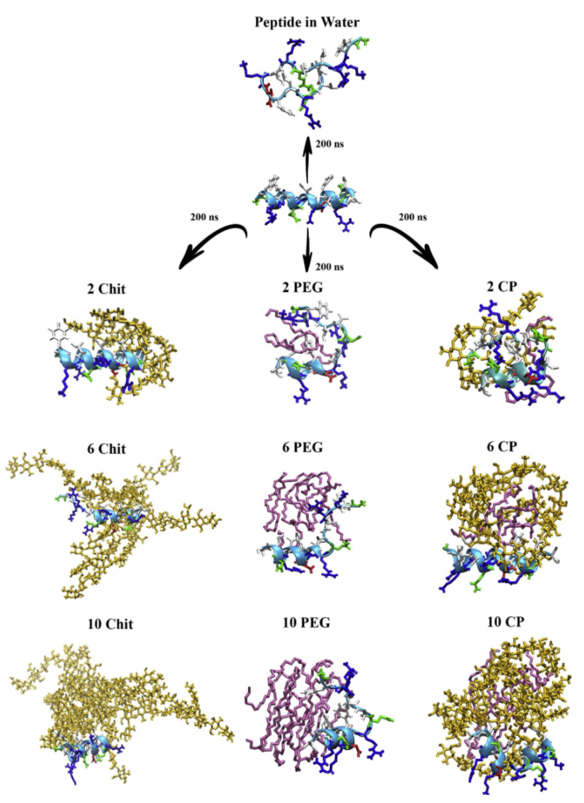
Wu et al. performed all-atom simulations of cecropin P1 grafted to the silica surface via a PEG cross-linker, showing the effect of PEG size and ionic strength on the conformation and antimicrobial activity of the peptide [79,80]. Our group simulated PEGylated magainin 2 and tachyplesin I interacting with lipid bilayers, showing that PEGylation reduces the binding strength between peptides and bilayer surfaces, which occurs more significantly for α-helical magainin 2 than for β-sheet tachyplesin I [81]. Recently, Jafari et al. simulated PEG-encapsulated magainin 2 and found that the PEG–peptide interaction is significantly modulated by aromatic and basic residues of the peptide [82], and Souza et al. simulated the insertion of PEG-encapsulated human beta-defensin-3 to lung surfactant models, showing that PEG chains promote the translocation of the peptide from gas phase to water phase [83]. Asadzadeh et al. simulated GF-17 (17th–32nd residues of LL-37) interacting with chitosan, PEG, or both, showing that the peptide interacts more tightly with PEG than with chitosan (Figure 3), leading to lower helicity in the presence of PEG [84].
2.3. Coiled Coil Peptides
Coiled coils are peptides composed of two or more α-helices wound into a superhelix. Sequences of coiled coils contain a heptad repeat of seven amino acid residues, where the 1st and 4th residues of each heptad repeat are hydrophobic [85]. These hydrophobic residues are located in the core of coiled coils and thus stabilize the superhelical structure of coiled coil bundles [85]. Coiled coils are found in approximately 10% of all proteins and serve critical roles as mediators of oligomerization of many proteins such as transcription factors, molecular motors, receptors and signaling molecules [86]. In addition, coiled coils can self-assemble to mechanically rigid protein fibers and thus have been synthesized for drug delivery applications as templates to promote the assembly of other molecules [87,88]. To increase their solubility and stability, PEGylated coiled coils have been experimentally synthesized by the Klok group [89,90,91,92,93], the Kros group [94,95,96,97,98,99,100,101,102,103], and the Xu group [104,105,106,107,108,109,110,111,112,113], showing membrane fusion and micelle assembly modulated by peptide sequence, PEG size and density, which have been theoretically complemented by simulations.
Jain and Ashbaugh performed replica exchange simulations of PEGylated coiled coils, showing the higher helicity of coiled coils grafted with larger PEG chains due to the interactions between PEG and lysine residues of peptides [114]. The Keten group found strong interactions of PEG chains with both hydrophobic and polar residues of peptides, leading to increased helicity and decreased solvent-accessible surface area of the peptide in the presence of PEG, to an extent dependent on solvent hydrophobicity [115]. The binding energy of a cyclic peptide dimer was influenced by PEG length and grafting density [116]. They also found the high helicity and stability of coiled coils grafted with low-molecular weight PEG chains [117], and the extended structure of PEG chains grafted to helix micelles [110]. In particular, PEGylation influences the micelle size and stability, which is interpreted by a competition between the entropy of PEG conformations in the assembled state [111,118]. Recently, they performed both all-atom and CG simulations of PEGylated coiled coils composed of three or four helices, showing the formation of self-assembled micelles and the effect of the coiled coil oligomeric state on micelle size and stability [119]. Our group also performed all-atom and CG simulations of PEGylated trimeric coiled coils and their self-assembled micelles (Figure 4), and calculated their radii of gyration and hydrodynamic radii, which favorably compare with experimental values [120]. In particular, we found that hydrophobic residues in the exterior sites of coiled coils tend to be less exposed to water and thus interact with PEG, leading to the compact conformation of adsorbed PEG [120].
3. PEGylated Liposomes
Liposomes, which are synthetic vesicles composed of phospholipid membranes, can transport drug molecules across specific cell membranes and thus have been widely used for drug delivery applications [121,122,123]. To increase solubility and circulating time of drug-encapsulating liposomes, PEG has been often attached to the liposome surface [2], since the first PEGylated liposomal doxorubicin (hydrophobic anticancer drug) was approved by the FDA in 1995 [124]. As the PEG size and concentration increase, encapsulated drug molecules can be more safely shielded from plasma proteins in the bloodstream, but also liposome membranes become unstable [125,126]. Therefore, many experiments have been performed to determine the maximum size and grafting density of PEG that can still maintain liposome stability [125,127,128,129,130,131,132,133,134,135,136,137,138,139,140], which has motivated theoretical studies on the effect of PEG size and grafting density on the conformation and dynamics of PEG chains grafted on the surface.
The Alexander–de Gennes theory has been applied to predict the transition of hydrophilic polymer chains between hemisphere (mushroom) and brush-like states on the surface [141]. Briefly, at very low grafting density, the grafted chain behaves like an isolated chain in solution, leading to a hemisphere (mushroom) conformation with a size given by the Flory radius, RF = aN3/5, where N is the degree of polymerization and a is the monomer size (Figure 5). At high grafting density (D < RF), polymer chains become crowded and repel each other, leading to a brush-like conformation with a thickness given by L = Na(a/D)2/3, where D is the distance between the grafting points of polymers. Jeon et al. calculated free energies of steric repulsion, van der Waals attraction, and hydrophobic interaction for the binding between spherical model proteins and PEO chains grafted on the hydrophobic surface, to an extent dependent on PEO length and grafting density [142]. Their free-energy calculations show that longer size and higher density (i.e., the brush state) lead to the optimal protein resistance, although surface density is more influential than chain length [142]. In particular, they determined optimal grafting densities of PEO for differently sized proteins, which was interpreted by steric repulsion and hydrophobic interaction between protein and PEO layer [143]. Szleifer also calculated free energies and showed the dependence of protein adsorption on the PEG density as well as on the protein conformation and concentration [144]. Halperin found that adsorption of small proteins can be repressed by increasing the grafting density, while adsorption of large proteins can be suppressed by increasing the brush thickness [145]. They also distinguish specific and nonspecific attractive interactions between various plasma proteins and PEG brushes [146]. Taylor and Jones found that the amount of proteins adsorbed onto PEGylated gold surfaces exponentially decreases as the brush density increases [147].
To complement these theoretical models, MD simulations have been performed. The Roccatano group performed all-atom simulations of PEGylated lipid bilayers, and their free-energy calculations showed the strong interactions between PEG and lipid headgroups of bilayers [148,149]. Bunker and coworkers parameterized the all-atom PEG model and simulated PEGylated lipid bilayers, showing the interactions between PEG oxygens and Na+ ions, and the penetration of PEG chains into a liquid-crystalline membrane but not into a gel-phased membrane [16]. They also found that the strength of the interaction between PEG and salt is modulated by PEG density, salt concentration and type such as NaCl, KCl, and CaCl2 [150]. They simulated small peptides interacting with PEGylated lipid bilayers, showing the dependence of peptide penetration on hydrophobicity [151]. In particular, Na+ ions bind to lipid bilayers and PEG chains grafted to drug molecules, which induces electrostatic repulsive interactions between lipid bilayers and PEGylated drugs [152]. PEGylation modulates the effect of cholesterol on the conformation and dynamics of lipid bilayers [153]. Their simulations also captured the insertion of hydrophobic drug or light-sensitizing molecules (e.g., porphyrins, indocyanine green, itraconazole, and piroxicam) to the PEG layer and the hydrophobic region of the bilayer (Figure 6) [154,155,156,157,158]. Recently, they simulated linear and branched PEG chains grafted on lipid bilayers, showing that the architecture and length of PEG–lipid conjugates influence the structure and dynamics of membranes, in agreement with experimental results [159].
Although all-atom simulations have captured the conformation and dynamics of PEGylated bilayers and their interactions with hydrophobic drug molecules and salt ions, the effects of PEG size and grafting density on liposome formation and protein adsorption have not been systematically simulated due to computational limitations of system size and time scale. To resolve this, the Klein group parameterized the CG model for PEG and PEGylated surfactants [160] and investigated the interactions between PEGylated surfactants and lipid bilayers [161] and the conformation of self-assembled PEGylated bicelles [162]. Our group also developed CG PEG model within the framework of the MARTINI FF [163,164], which lumps a monomer of PEG (–(CH2–CH2–O)n–; three heavy atoms) into each CG bead [18,25]. This CG PEG model was further parameterized to increase dihedral stability by Rossi et al., showing the effect of PEGylation on the curvature of the surfactant bilayer [26]. Using this CG PEG model, Yang and Faller found that the presence of PEGylated lipid promotes the conformational transition from bilayers to micelles [165]. Hezaveh et al. developed another version of the MARTINI-based PEG model and showed the insertion of block copolymers into lipid bilayers, although their model does not include a dihedral potential and thus cannot reproduce the experimentally observed conformation of PEG chains in water [166].
Our group simulated a mixture of lipids and PEGylated lipids at different molar ratios, showing the formation of self-assembled liposomes, bicelles, and micelles, respectively, at 0–2.2, 10.5–27.4, and higher mol% of PEGylated lipid, in qualitative agreement with experiments [167]. This indicates that the phase behavior and size of lipid assemblies can be modulated by PEG density because their bulky headgroups increase membrane curvature [167]. Moreover, our CG simulations of PEG chains grafted to a nonadsorbing surface captured the conformational transition between brush and mushroom states, showing good agreement of simulation and Alexander–de Gennes theory [25]. In particular, we characterized the extent of protein adsorption to PEGylated lipid bilayers in terms of different PEG sizes (Mw = 750, 2000, and 5000) and grafting densities (1.6–25 mol%), showing that the binding between proteins and membranes is suppressed by the PEG layer in a brush but not in a mushroom (Figure 7), in quantitative agreement with the Alexander–de Gennes theory and experiments regarding much less adsorption of plasma proteins onto the membrane surface grafted with PEG in the brush state than in the mushroom state [168]. It is worth noting that the binding between plasma protein and bilayer surface can be predicted from the boundary between mushroom and brush states of PEG with different sizes and grafting densities, as highlighted in Figure 7. Recently, Sammalkorpi and coworkers showed the formation of self-assembled liposomes, bicelles, and micelles at different PEGylated-lipid concentrations [169,170], similar to our previous work [167].
4. PEGylated Nanoparticles
4.1. Dendrimers
Dendrimers, which consist of regularly branched monomeric building blocks with many surface terminal groups, have shown great potential for drug delivery applications because of their controlled mass, surface valency, and surface functionality [171]. Drug molecules can be either conjugated to the terminal group of dendrimer or encapsulated into the inner vacancy of dendrimer and then delivered to the desired site [172]. However, charged dendrimers have nonspecific interactions with cell membranes and thus have been neutralized by acetylating their surface terminals. In addition, PEG chains have been often attached to the dendrimer surface, which does not only decrease nonspecific cytotoxicity but also increases dendrimer solubility [173]. In particular, PEG can sterically shield drug molecules from plasma proteins in the bloodstream and thus increase their circulation lifetime [174], which has motivated many simulation studies on the conformation of PEGylated dendrimers and their interaction with drugs, proteins, and lipid membranes.
Tanis and Karatasos performed all-atom simulations of a dendrimer grafted with a single PEO chain, showing the effect of pH on the conformation of PEO and its hydrogen-bond interaction with dendrimer [175]. Karatosas also simulated the complex of PEGylated hyperbranched polyesters and doxorubicin, showing the effects of PEG size and doxorubicin charge on the hydrogen-bond interactions between PEGylated polyesters and doxorubicin [176]. Our CG simulations showed that PEGylation induces interparticle dispersion [177] and the lower extent of cytotoxicity and membrane permeability than acetylation does [178]. In particular, we found that longer chains with higher grafting densities promote PEG–PEG crowding and thus stretch dendrimer terminals towards water, leading to a larger dendrimer with a dense-shell structure [179]. Albertazzi et al. simulated dendrimers containing 2- and 4-arm PEG cores, showing more swollen conformation of dendrimer at higher concentrations of PEG core [180]. Their metadynamics simulations also showed that PEGylated dendrimers have a tight globular shape rather than an open conformation [181]. Pearson et al. showed conformational changes of PEGylated dendrimers at different charge densities [182], and Lin et al. found that PEGylated dendrimers adsorb to lipid monolayers but do not significantly influence the structure and properties of monolayers [183].
Recently, large complexes of PEGylated dendrimers and proteins (or drugs) have been simulated. Lim et al. [184] and Barraza et al. [185] respectively simulated paclitaxel and methotrexate drugs interacting PEGylated dendrimers, showing that PEG–PEG crowding decreases the extent of drug release, which helps determine the size and density of PEG for optimal drug release. Sampogna-Mireles et al. simulated dendrimers grafted with PEG and folic acid, showing that PEG chains do not reduce the binding affinity between folic acid and folate receptor (Figure 8) [186], which should be highlighted because their simulations captured the effect of PEG on the binding affinity to the receptor protein. Hsu et al. simulated PEGylated dendron micelle and serum albumin, showing that the penetration of serum albumin into the micelle core can be suppressed by PEGylation [187]. Diaz et al. compared the conformations of dendrimers grafted with PEG or folic acid, showing different effects of PEG and folic acid on dendrimer size, which helps explain the experimentally observed relationship between dendrimer size and circulation time [188]. Wang et al. found that PEGylation significantly weakens the binding between dendrimers and plasma proteins such as human serum albumin and immunoglobulin [189]. Overall, the conformation of PEG and its effect on the internal structure of dendrimer were mainly studied until early 2010s, while the effect of PEGylation on the binding affinity to proteins and drug release efficiency have been more focused for the past five years.
4.2. Carbon Nanotubes
Since CNTs are mechanically strong and chemically stable, they have been considered to be good candidate nanomaterials for use as drug transporters [190,191,192]. However, CNTs are highly hydrophobic and thus immediately aggregate in aqueous environment, which limits the application of CNTs at the physiological condition. To overcome this, PEG chains have been covalently or noncovalently attached to the CNT surface. Experiments have revealed the conformation and interparticle dispersion of PEGylated CNTs and their interactions with membranes, proteins, and drug molecules [193,194,195,196,197,198,199,200], which has motivated simulation studies.
In the early 2010s, most simulation studies focused on the conformation of PEG chains and their interactions with the CNT surface. Uddin et al. performed all-atom simulations of CNTs with a mixture of PEO and water, and their free-energy calculations showed the weak adsorption of PEO onto the CNT surface, which were explained by entropic and enthalpic contributions [201]. Our group simulated random adsorption of PEGylated lipids onto the CNT surface [202] and found the effects of PEG size and grafting density on the conformation of PEG grafted onto CNT [203], which favorably compares with the transition of mushroom and brush states in the Alexander–de Gennes theory [141]. Di Crescenzo et al. simulated CNTs interacting with PEG (homopolymer) or PEG–propylene sulfide (PPS) block copolymers and found the stronger interaction of CNT with PEG–PPS than with PEG and the parallel arrangement PEG chains along the tube axis [204]. Aslan et al. compares density profiles of PEGylated lipids adsorbed onto isolated and bundled CNTs, which helps explain their different extents of antimicrobial activity [205]. Sarukhanyan et al. [206] and Han et al. [207] simulated CNTs interacting with various polymers, showing the effect of polymer hydrophobicity on the CNT-polymer conformation and interparticle dispersion. Maatta et al. found the dependence of CNT dispersion on PEG length and CNT diameter [208].
Recently, large complexes of PEGylated CNTs and other molecules such as lipid membranes, plasma proteins, and anticancer drug molecules have been simulated using all-atom and CG models. Skandani and Al-Haik showed slower penetration of PEGylated CNT into the lipid bilayer than unPEGylated CNT, which was explained by lower adhesion energy of PEGylated CNT [209], as observed in their previous experiments [210]. Our group showed interparticle dispersion and membrane curvature induced by PEGylated CNT [211], and the effects of protein shape, PEG size and grafting density on the adsorption of proteins onto PEGylated CNT [212]. Lin et al. investigated the binding affinity between CNTs and PEGylated proteins such as hormones, neurotransmitter, and vitamin [213], and Hashemzadeh and Raissi showed the adsorption of paclitaxel onto the PEGylated CNT [214]. Kavyani et al. compared the binding strength of CNTs with PEGylated and unPEGylated dendrimers, showing the stronger interactions of CNTs with PEGylated dendrimers than with unPEGylated dendrimers [215]. The Panczyk group performed all-atom simulations of PEGylated and folic acid-functionalized CNTs that encapsulate doxorubicin, showing the release of doxorubicin from CNTs at acidic pH but not at neutral pH [216]. Fullerene molecules were also included to the inner cavity of CNT functionalized with PEG and folic acid, where fullerenes act as magnetic pistons at acidic pH, leading to an increase in the release of doxorubicin from nanotube [217], which helps explain the effect of PEG on the efficiency of drug release as well as suggests the use of fullerene, as presented in Figure 9. Meran et al. simulated CNTs coated with PEGylated pyrene and showed the adsorption of PEGylated pyrene onto the CNT surface via π-π stacking interactions, which does not significantly depend on PEG length and CNT size [218]. Saberinasab et al. performed quantum-mechanics (QM) calculations and all-atom MD simulations of a mixture of PEGylated CNTs and Temozolomide (anticancer drug), showing the adsorption of Temozolomide on PEGylated CNT because of strong hydrogen-bond interactions [219]. Moradnia et al. also performed QM calculations and all-atom MD simulations of a mixture of PEGylated CNTs and Gemcitabine (anticancer drug), showing the effect of PEG size on the hydrogen-bond interactions of Gemcitabine with PEGylated CNT and water [220]. Overall, simulation studies focused on the conformation of PEG and its effect on CNT dispersion until the mid-2010s, but have recently focused more closely on the effect of PEGylation on the efficiency of drug release and the binding affinity to drugs and proteins.
5. Conclusions
All-atom and CG MD simulations have revealed much useful information about the structure and dynamics of PEGylated drug transporters such as proteins, peptides, liposomes, dendrimers, and CNTs, which cannot be easily captured by experiments. In 1995–2000s, all-atom and CG PEG FFs have been developed and used for simulations of short linear PEG and their interactions with small molecules and solvents, while recent advances in computer power and simulation methods have allowed simulations of large complexes of PEGylated drug carries and their interactions with anticancer drugs, plasma proteins, lipid membranes, and receptors.
Although the molecular simulation has proven to be a powerful tool for the in silico design of PEGylated drugs for the past two decades, there are still problems that need to be considered for the future work. Firstly, biological complexes, reaction kinetics and mass transport conditions of experiments and simulations differ, which precludes any quantitative comparison between simulations and experiments. For instance, there are hundreds of plasma proteins that flow through the bloodstream, and hundreds of membrane proteins that control cellular behavior and interactions with drug carriers. Recently, MD simulations start to simulate the corona formation of various plasma proteins [221] and human cell membranes composed of 63 different lipid species [222], showing promising efforts in mimics of biological systems. Secondly, more accurate FFs need to be developed to predict the interactions between PEG and other molecules such as nucleotides and amino acids. As reviewed above, all-atom and CG PEG FFs have accurately predicted the conformation and physical properties of PEG in solvent, but the prediction of their interactions with other molecules need to be improved. Lastly, large complexes of PEGylated drugs interacting with other molecules should be considered. This can be done by multiscale simulations of the transition between all-atom and CG models [223], where the conformation and dynamics can be equilibrated by CG simulations, and then CG coordinates can be converted to all-atom models that offer insights into the atomic-level interactions such as electrostatic, hydrophobic, and hydrogen-bond interactions. To achieve this, simulation methodologies need to be developed for the transition from CG to an all-atom model that can be compatible with various biomolecules, polymers, surfactants, and solvents.
Despite these limitations, MD simulations have successfully interpreted experimental observations at nearly the atomic scale and determined the optimal size, structure, and grafting density of PEG. Moreover, an increase in computational speed and methodology development (e.g., multiscale simulations of the transition between all-atom and CG models) will allow for more realistic simulations of larger biological systems, leading to a promising tool for the rational design of highly efficient PEGylated drug delivery systems.
Funding
This research received no external funding.
Conflicts of Interest
The authors declare no conflict of interest.
References
- Harris, J.M.; Chess, R.B. Effect of pegylation on pharmaceuticals. Nat. Rev. Drug Discov. 2003, 2, 214–221. [Google Scholar] [CrossRef]
- Harris, J.M.; Martin, N.E.; Modi, M. Pegylation—A novel process for modifying pharmacokinetics. Clin. Pharmacokinet. 2001, 40, 539–551. [Google Scholar] [CrossRef]
- Veronese, F.M.; Pasut, G. PEGylation, successful approach to drug delivery. Drug Discov. Today 2005, 10, 1451–1458. [Google Scholar] [CrossRef]
- Allen, T.M.; Hansen, C. Pharmacokinetics of Stealth Versus Conventional Liposomes—Effect of Dose. Biochim. Biophys. Acta 1991, 1068, 133–141. [Google Scholar] [CrossRef]
- Allen, T.M.; Hansen, C.; Martin, F.; Redemann, C.; Yauyoung, A. Liposomes Containing Synthetic Lipid Derivatives of Poly(Ethylene Glycol) Show Prolonged Circulation Half-Lives Invivo. Biochim. Biophys. Acta 1991, 1066, 29–36. [Google Scholar] [CrossRef]
- Maruyama, K.; Yuda, T.; Okamoto, A.; Ishikura, C.; Kojima, S.; Iwatsuru, M. Effect of Molecular-Weight in Amphipathic Polyethyleneglycol on Prolonging the Circulation Time of Large Unilamellar Liposomes. Chem. Pharm. Bull. 1991, 39, 1620–1622. [Google Scholar] [CrossRef] [PubMed] [Green Version]
- Delgado, C.; Francis, G.E.; Fisher, D. The uses and properties of peg-linked proteins. Crit. Rev. Ther. Drug Carr. Syst. 1992, 9, 249–304. [Google Scholar]
- Maruyama, K.; Yuda, T.; Okamoto, A.; Kojima, S.; Suginaka, A.; Iwatsuru, M. Prolonged circulation time invivo of large unilamellar liposomes composed of distearoyl phosphatidylcholine and cholesterol containing amphipathic poly(ethylene glycol). Biochim. Biophys. Acta 1992, 1128, 44–49. [Google Scholar] [CrossRef]
- Duncan, R. Polymer conjugates as anticancer nanomedicines. Nat. Rev. Cancer 2006, 6, 688–701. [Google Scholar] [CrossRef] [PubMed]
- Turecek, P.L.; Bossard, M.J.; Schoetens, F.; Ivens, I.A. PEGylation of Biopharmaceuticals: A Review of Chemistry and Nonclinical Safety Information of Approved Drugs. J. Pharm. Sci. 2016, 105, 460–475. [Google Scholar] [CrossRef] [Green Version]
- Knop, K.; Hoogenboom, R.; Fischer, D.; Schubert, U.S. Poly(ethylene glycol) in drug delivery: Pros and cons as well as potential alternatives. Angew. Chem. Int. Ed. 2010, 49, 6288–6308. [Google Scholar] [CrossRef] [PubMed]
- Sousa, S.F.; Peres, J.; Coelho, M.; Vieira, T.F. Analyzing PEGylation through Molecular Dynamics Simulations. ChemistrySelect 2018, 3, 8415–8427. [Google Scholar] [CrossRef]
- Rossi, G.; Monticelli, L. Modeling the effect of nano-sized polymer particles on the properties of lipid membranes. J. Phys. Condens. Matter 2014, 26, 503101. [Google Scholar] [CrossRef] [PubMed]
- Lee, H. Molecular modeling of PEGylated peptides, dendrimers, and single-walled carbon nanotubes for biomedical applications. Polymers 2014, 6, 776–798. [Google Scholar] [CrossRef] [Green Version]
- Bedrov, D.; Borodin, O.; Smith, G.D. Molecular dynamics simulations of 1,2-dimethoxyethane/water solutions. 1. Conformational and structural properties. J. Phys. Chem. B 1998, 102, 5683–5690. [Google Scholar] [CrossRef]
- Stepniewski, M.; Pasenkiewicz-Gierula, M.; Rog, T.; Danne, R.; Orlowski, A.; Karttunen, M.; Urtti, A.; Yliperttula, M.; Vuorimaa, E.; Bunker, A. Study of PEGylated lipid layers as a model for PEGylated liposome surfaces: Molecular dynamics simulation and langmuir monolayer studies. Langmuir 2011, 27, 7788–7798. [Google Scholar] [CrossRef] [PubMed]
- Vorobyov, I.; Anisimov, V.M.; Greene, S.; Venable, R.M.; Moser, A.; Pastor, R.W.; MacKerell, A.D. Additive and classical drude polarizable force fields for linear and cyclic ethers. J. Chem. Theory Comput. 2007, 3, 1120–1133. [Google Scholar] [CrossRef] [PubMed]
- Lee, H.; Venable, R.M.; MacKerell, A.D.; Pastor, R.W. Molecular dynamics studies of polyethylene oxide and polyethylene glycol: Hydrodynamic radius and shape anisotropy. Biophys. J. 2008, 95, 1590–1599. [Google Scholar] [CrossRef] [PubMed] [Green Version]
- Dong, H.; Hyun, J.K.; Durham, C.; Wheeler, R.A. Molecular dynamics simulations and structural comparisons of amorphous poly(ethylene oxide) and poly(ethylenimine) models. Polymer 2001, 42, 7809–7817. [Google Scholar] [CrossRef]
- Tritopoulou, E.A.; Economou, I.G. Molecular simulation of structure and thermodynamic properties of pure tri- and tetra-ethylene glycols and their aqueous mixtures. Fluid Phase Equilibria 2006, 248, 134–146. [Google Scholar] [CrossRef]
- Winger, M.; De Vries, A.H.; Van Gunsteren, W.F. Force-field dependence of the conformational properties of α, ω-dimethoxypolyethylene glycol. Mol. Phys. 2009, 107, 1313–1321. [Google Scholar] [CrossRef] [Green Version]
- Neyertz, S.; Brown, D.; Thomas, J.O. Molecular dynamics simulation of crystalline poly(ethylene oxide). J. Chem. Phys. 1994, 101, 10064–10073. [Google Scholar] [CrossRef]
- Lin, B.; Boinske, P.T.; Halley, J.W. A molecular dynamics model of the amorphous regions of polyethylene oxide. J. Chem. Phys. 1996, 105, 1668–1681. [Google Scholar] [CrossRef]
- Srinivas, G.; Discher, D.E.; Klein, M.L. Self-assembly and properties of diblock copolymers by coarse-grain molecular dynamics. Nat. Mater. 2004, 3, 638–644. [Google Scholar] [CrossRef] [PubMed]
- Lee, H.; de Vries, A.H.; Marrink, S.J.; Pastor, R.W. A Coarse-Grained Model for Polyethylene Oxide and Polyethylene Glycol: Conformation and Hydrodynamics. J. Phys. Chem. B 2009, 113, 13186–13194. [Google Scholar] [CrossRef] [Green Version]
- Rossi, G.; Fuchs, P.F.J.; Barnoud, J.; Monticelli, L. A coarse-grained MARTINI model of polyethylene glycol and of polyoxyethylene alkyl ether surfactants. J. Phys. Chem. B 2012, 116, 14353–14362. [Google Scholar] [CrossRef]
- Choi, E.; Mondal, J.; Yethiraj, A. Coarse-grained models for aqueous polyethylene glycol solutions. J. Phys. Chem. B 2014, 118, 323–329. [Google Scholar] [CrossRef]
- Wang, Q.; Keffer, D.J.; Nicholson, D.M. A coarse-grained model for polyethylene glycol polymer. J. Chem. Phys. 2011, 135. [Google Scholar] [CrossRef] [PubMed]
- Chen, T.; Hynninen, A.P.; Prud’homme, R.K.; Kevrekidis, I.G.; Panagiotopoulos, A.Z. Coarse-Grained Simulations of Rapid Assembly Kinetics for Polystyrene-b-poly(ethylene oxide) Copolymers in Aqueous Solutions. J. Phys. Chem. B 2008, 112, 16357–16366. [Google Scholar] [CrossRef]
- Fischer, J.; Paschek, D.; Geiger, A.; Sadowski, G. Modeling of Aqueous Poly(oxyethylene) Solutions. 2. Mesoscale Simulations. J. Phys. Chem. B 2008, 112, 13561–13571. [Google Scholar] [CrossRef] [PubMed]
- Bedrov, D.; Ayyagari, C.; Smith, G.D. Multiscale modeling of poly(ethylene oxide)-poly(propylene oxide)-poly(ethylene oxide) triblock copolymer micelles in aqueous solution. J. Chem. Theory Comput. 2006, 2, 598–606. [Google Scholar] [CrossRef] [PubMed]
- Abuchowski, A.; McCoy, J.R.; Palczuk, N.C.; van Es, T.; Davis, F.F. Effect of covalent attachment of polyethylene glycol on immunogenicity and circulating life of bovine liver catalase. J. Biol. Chem. 1977, 252, 3582–3586. [Google Scholar] [PubMed]
- Abuchowski, A.; Van Es, T.; Palczuk, N.C.; Davis, F.F. Alteration of immunological properties of bovine serum albumin by covalent attachment of polyethylene glycol. J. Biol. Chem. 1977, 252, 3578–3581. [Google Scholar] [PubMed]
- Piedmonte, D.M.; Treuheit, M.J. Formulation of Neulasta® (pegfilgrastim). Adv. Drug Deliv. Rev. 2008, 60, 50–58. [Google Scholar] [CrossRef] [PubMed]
- Ettinger, A.R. Pegaspargase (Oncaspar). J. Pediatric Oncol. Nurs. 1995, 12, 46–48. [Google Scholar] [CrossRef] [PubMed]
- Lang, L. FDA Approves Cimzia to Treat Crohn’s Disease. Gastroenterology 2008, 134, 1819. [Google Scholar] [CrossRef] [PubMed]
- Sánchez-Fructuoso, A.; Guirado, L.; Ruiz, J.C.; Torregrosa, V.; Gonzalez, E.; Suarez, M.L.; Galléego, R. Anemia control in kidney transplant patients treated with methoxy polyethylene glycol-epoetin beta (Mircera): The anemiatrans group. Transplant. Proc. 2010, 42, 2931–2934. [Google Scholar] [CrossRef]
- Parfrey, P.S.; Warden, G.; Barrett, B.J. On peginesatide and anemia treatment in CKD. Am. J. Kidney Dis. 2013, 62, 659–661. [Google Scholar] [CrossRef]
- Shukla, D.; Namperumalsamy, P.; Goldbaum, M.; Cunningham, E.T., Jr. Pegaptanib sodium for ocular vascular disease. Indian J. Ophthalmol. 2007, 55, 427–430. [Google Scholar] [PubMed]
- Hoy, S.M. Peginterferon beta-1a: A review of its use in patients with relapsing-remitting multiple sclerosis. CNS Drugs 2015, 29, 171–179. [Google Scholar] [CrossRef] [PubMed]
- Berhanu, A.A.; Krasnokutsky, S.; Keenan, R.T.; Pillinger, M.H. Pegloticase failure and a possible solution: Immunosuppression to prevent intolerance and inefficacy in patients with gout. Semin. Arthritis Rheum. 2017, 46, 754–758. [Google Scholar] [CrossRef] [PubMed]
- Pasut, G.; Veronese, F.M. Polymer-drug conjugation, recent achievements and general strategies. Prog. Polym. Sci. 2007, 32, 933–961. [Google Scholar] [CrossRef]
- Jevševar, S.; Kunstelj, M.; Porekar, V.G. PEGylation of therapeutic proteins. Biotechnol. J. 2010, 5, 113–128. [Google Scholar] [CrossRef] [PubMed] [Green Version]
- Burdick, J.A.; Anseth, K.S. Photoencapsulation of osteoblasts in injectable RGD-modified PEG hydrogels for bone tissue engineering. Biomaterials 2002, 23, 4315–4323. [Google Scholar] [CrossRef]
- Roberts, M.J.; Bentley, M.D.; Harris, J.M. Chemistry for peptide and protein PEGylation. Adv. Drug Deliv. Rev. 2002, 54, 459–476. [Google Scholar] [CrossRef]
- Caliceti, P.; Veronese, F.M. Pharmacokinetic and biodistribution properties of poly(ethylene glycol)-protein conjugates. Adv. Drug Deliv. Rev. 2003, 55, 1261–1277. [Google Scholar] [CrossRef]
- Manjula, B.N.; Tsai, A.; Upadhya, R.; Perumalsamy, K.; Smith, P.K.; Malavalli, A.; Vandegriff, K.; Winslow, R.M.; Intaglietta, M.; Prabhakaran, M.; et al. Site-specific PEGylation of hemoglobin at Cys-93(β): Correlation between the colligative properties of the PEGylated protein and the length of the conjugated PEG chain. Bioconjugate Chem. 2003, 14, 464–472. [Google Scholar] [CrossRef] [PubMed]
- Meng, F.; Manjula, B.N.; Tsai, A.G.; Cabrales, P.; Intaglietta, M.; Smith, P.K.; Prabhakaran, M.; Acharya, S.A. Hexa-thiocarbamoyl phenyl PEG5K Hb: Vasoactivity and structure: The conjugation linkage on the pegylation induced plasma expander-like solution properties of PEG-Hb adducts. Protein J. 2009, 28, 199–212. [Google Scholar] [CrossRef] [PubMed]
- Yang, C.; Lu, D.; Liu, Z. How PEGylation Enhances the Stability and Potency of Insulin: A Molecular Dynamics Simulation. Biochemistry 2011, 50, 2585–2593. [Google Scholar] [CrossRef]
- Zhang, Y.; Han, K.; Lu, D.; Liu, Z. Reversible encapsulation of lysozyme within mPEG-b-PMAA: Experimental observation and molecular dynamics simulation. Soft Matter 2013, 9, 8723–8729. [Google Scholar] [CrossRef]
- Chen, G.; Kong, X.; Zhu, J.; Lu, D.; Liu, Z. How ABA block polymers activate cytochrome c in toluene: Molecular dynamics simulation and experimental observation. Phys. Chem. Chem. Phys. 2015, 17, 10708–10714. [Google Scholar] [CrossRef] [PubMed]
- Xue, Y.; O’Mara, M.L.; Surawski, P.P.T.; Trau, M.; Mark, A.E. Effect of poly(ethylene glycol) (PEG) spacers on the conformational properties of small peptides: A molecular dynamics study. Langmuir 2011, 27, 296–303. [Google Scholar] [CrossRef] [PubMed]
- Mu, Q.; Hu, T.; Yu, J. Molecular Insight into the Steric Shielding Effect of PEG on the Conjugated Staphylokinase: Biochemical Characterization and Molecular Dynamics Simulation. PLoS ONE 2013, 8, e68559. [Google Scholar] [CrossRef] [PubMed] [Green Version]
- Khameneh, B.; Jaafari, M.R.; Hassanzadeh-Khayyat, M.; Varasteh, A.; Chamani, J.; Iranshahi, M.; Mohammadpanah, H.; Abnous, K.; Saberi, M.R. Preparation, characterization and molecular modeling of PEGylated human growth hormone with agonist activity. Int. J. Biol. Macromol. 2015, 80, 400–409. [Google Scholar] [CrossRef] [PubMed]
- Diaferia, C.; Sibillano, T.; Balasco, N.; Giannini, C.; Roviello, V.; Vitagliano, L.; Morelli, G.; Accardo, A. Hierarchical Analysis of Self-Assembled PEGylated Hexaphenylalanine Photoluminescent Nanostructures. Chem. A Eur. J. 2016, 22, 16586–16597. [Google Scholar] [CrossRef] [PubMed] [Green Version]
- Diaferia, C.; Balasco, N.; Altamura, D.; Sibillano, T.; Gallo, E.; Roviello, V.; Giannini, C.; Morelli, G.; Vitagliano, L.; Accardo, A. Assembly modes of hexaphenylalanine variants as function of the charge states of their terminal ends. Soft Matter 2018, 14, 8219–8230. [Google Scholar] [CrossRef] [PubMed]
- Diaferia, C.; Balasco, N.; Sibillano, T.; Giannini, C.; Vitagliano, L.; Morelli, G.; Accardo, A. Structural Characterization of Self-Assembled Tetra-Tryptophan Based Nanostructures: Variations on a Common Theme. ChemPhysChem 2018, 19, 1635–1642. [Google Scholar] [CrossRef]
- Diaferia, C.; Balasco, N.; Sibillano, T.; Ghosh, M.; Adler-Abramovich, L.; Giannini, C.; Vitagliano, L.; Morelli, G.; Accardo, A. Amyloid-Like Fibrillary Morphology Originated by Tyrosine-Containing Aromatic Hexapeptides. Chem. A Eur. J. 2018, 24, 6804–6817. [Google Scholar] [CrossRef] [PubMed]
- Settanni, G.; Zhou, J.; Schmid, F. Interactions between proteins and poly(ethylene-glycol) investigated using molecular dynamics simulations. Proc. J. Phys. Conf. Ser. 2017, 921, 012002. [Google Scholar] [CrossRef]
- Settanni, G.; Zhou, J.; Suo, T.; Schöttler, S.; Landfester, K.; Schmid, F.; Mailänder, V. Protein corona composition of poly(ethylene glycol)-and poly (phosphoester)-coated nanoparticles correlates strongly with the amino acid composition of the protein surface. Nanoscale 2017, 9, 2138–2144. [Google Scholar] [CrossRef] [PubMed] [Green Version]
- Settanni, G.; Schäfer, T.; Muhl, C.; Barz, M.; Schmid, F. Poly-sarcosine and Poly(Ethylene-Glycol) Interactions with Proteins Investigated Using Molecular Dynamics Simulations. Comput. Struct. Biotechnol. J. 2018, 16, 543–550. [Google Scholar] [CrossRef] [PubMed]
- Kurinomaru, T.; Kuwada, K.; Tomita, S.; Kameda, T.; Shiraki, K. Noncovalent PEGylation through Protein-Polyelectrolyte Interaction: Kinetic Experiment and Molecular Dynamics Simulation. J. Phys. Chem. B 2017, 121, 6785–6791. [Google Scholar] [CrossRef] [PubMed]
- Zaghmi, A.; Mendez-Villuendas, E.; Greschner, A.A.; Liu, J.Y.; de Haan, H.W.; Gauthier, M.A. Mechanisms of activity loss for a multi-PEGylated protein by experiment and simulation. Mater. Today Chem. 2019, 12, 121–131. [Google Scholar] [CrossRef]
- Sindhu, R.; Pradeep, H.; Manonmani, H.K. Polyethylene glycol acts as a mechanistic stabilizer of L-asparaginase: A computational probing. Med. Chem. 2019, 15, 703–711. [Google Scholar] [CrossRef] [PubMed]
- Ramezanghorbani, F.; Lin, P.; Colina, C.M. Optimizing Protein-Polymer Interactions in a Poly(ethylene glycol) Coarse-Grained Model. J. Phys. Chem. B 2018, 122, 7997–8005. [Google Scholar] [CrossRef]
- Munasinghe, A.; Mathavan, A.; Lin, P.; Colina, C.M. PEGylation within a confined hydrophobic cavity of a protein. Phys. Chem. Chem. Phys. 2019, 21, 25584–25596. [Google Scholar] [CrossRef]
- Baker, S.L.; Munasinghe, A.; Murata, H.; Lin, P.; Matyjaszewski, K.; Colina, C.M.; Russell, A.J. Intramolecular Interactions of Conjugated Polymers Mimic Molecular Chaperones to Stabilize Protein-Polymer Conjugates. Biomacromolecules 2018, 19, 3798–3813. [Google Scholar] [CrossRef]
- Rukmani, S.J.; Anstine, D.M.; Munasinghe, A.; Colina, C.M. An Insight into Structural and Mechanical Properties of Ideal-Networked Poly(Ethylene Glycol)–Peptide Hydrogels from Molecular Dynamics Simulations. Macromol. Chem. Phys. 2020, 221, 1900326. [Google Scholar] [CrossRef]
- Zasloff, M. Antimicrobial peptides of multicellular organisms. Nature 2002, 415, 389–395. [Google Scholar] [CrossRef] [PubMed]
- Matsuzaki, K. Why and how are peptide-lipid interactions utilized for self-defense? Magainins and tachyplesins as archetypes. Biochim. Biophys. Acta-Biomembr. 1999, 1462, 1–10. [Google Scholar] [CrossRef] [Green Version]
- Matsuzaki, K. Control of cell selectivity of antimicrobial peptides. Biochim. Biophys. Acta-Biomembr. 2009, 1788, 1687–1692. [Google Scholar] [CrossRef] [PubMed] [Green Version]
- Guiotto, A.; Pozzobon, M.; Canevari, M.; Manganelli, R.; Scarin, M.; Veronese, F.M. PEGylation of the antimicrobial peptide nisin A: Problems and perspectives. Farmaco 2003, 58, 45–50. [Google Scholar] [CrossRef]
- Imura, Y.; Nishida, M.; Matsuzaki, K. Action mechanism of PEGylated magainin 2 analogue peptide. Biochim. Biophys. Acta-Biomembr. 2007, 1768, 2578–2585. [Google Scholar] [CrossRef] [PubMed] [Green Version]
- Imura, Y.; Nishida, M.; Ogawa, Y.; Takakura, Y.; Matsuzaki, K. Action mechanism of tachyplesin I and effects of PEGylation. Biochim. Biophys. Acta-Biomembr. 2007, 1768, 1160–1169. [Google Scholar] [CrossRef] [PubMed] [Green Version]
- Singh, S.; Papareddy, P.; Mörgelin, M.; Schmidtchen, A.; Malmsten, M. Effects of PEGylation on Membrane and Lipopolysaccharide Interactions of Host Defense Peptides. Biomacromolecules 2014, 15, 1337–1345. [Google Scholar] [CrossRef] [PubMed]
- Gong, Y.; Andina, D.; Nahar, S.; Leroux, J.C.; Gauthier, M.A. Releasable and traceless PEGylation of arginine-rich antimicrobial peptides. Chem. Sci. 2017, 8, 4082–4086. [Google Scholar] [CrossRef] [PubMed] [Green Version]
- Morris, C.J.; Beck, K.; Fox, M.A.; Ulaeto, D.; Clark, G.C.; Gumbleton, M. Pegylation of antimicrobial peptides maintains the active peptide conformation, model membrane interactions, and antimicrobial activity while improving lung tissue biocompatibility following airway delivery. Antimicrob. Agents Chemother. 2012, 56, 3298–3308. [Google Scholar] [CrossRef] [Green Version]
- Falciani, C.; Lozzi, L.; Scali, S.; Brunetti, J.; Bracci, L.; Pini, A. Site-specific pegylation of an antimicrobial peptide increases resistance to Pseudomonas aeruginosa elastase. Amino Acids 2014, 46, 1403–1407. [Google Scholar] [CrossRef]
- Wu, X.; Chang, H.; Mello, C.; Nagarajan, R.; Narsimhan, G. Effect of interaction with coesite silica on the conformation of Cecropin P1 using explicit solvent molecular dynamics simulation. J. Chem. Phys. 2013, 138, 01B618. [Google Scholar] [CrossRef] [PubMed]
- Wu, X.; Wei, P.H.; Zhu, X.; Wirth, M.J.; Bhunia, A.; Narsimhan, G. Effect of immobilization on the antimicrobial activity of a cysteine-terminated antimicrobial Peptide Cecropin P1 tethered to silica nanoparticle against E. coli O157:H7 EDL933. Colloids Surf. B Biointerfaces 2017, 156, 305–312. [Google Scholar] [CrossRef] [PubMed]
- Han, E.; Lee, H. Effects of pegylation on the binding interaction of magainin 2 and tachyplesin I with lipid bilayer surface. Langmuir 2013, 29, 14214–14221. [Google Scholar] [CrossRef] [PubMed]
- Jafari, M.; Doustdar, F.; Mehrnejad, F. Molecular Self-Assembly Strategy for Encapsulation of an Amphipathic α-Helical Antimicrobial Peptide into the Different Polymeric and Copolymeric Nanoparticles. J. Chem. Inf. Modeling 2019, 59, 550–563. [Google Scholar] [CrossRef] [PubMed]
- Souza, F.R.; Souza, L.M.P.; Pimentel, A.S. Permeation of beta-defensin-3 encapsulated with polyethylene glycol in lung surfactant models at air-water interface. Colloids Surf. B Biointerfaces 2019, 182, 110357. [Google Scholar] [CrossRef] [PubMed]
- Asadzadeh, H.; Moosavi, A.; Arghavani, J.H. The effect of chitosan and PEG polymers on stabilization of GF-17 structure: A molecular dynamics study. Carbohydr. Polym. 2020, 237, 116124. [Google Scholar] [CrossRef] [PubMed]
- Lupas, A.N.; Gruber, M. The structure of alpha-helical coiled coils. In Fibrous Proteins: Coiled-Coils, Collagen and Elastomers; Elsevier Academic Press Inc.: San Diego, CA, USA, 2005; Volume 70, pp. 37–38. [Google Scholar]
- Gruber, M.; Lupas, A.N. Historical review: Another 50th anniversary-new periodicities in coiled coils. Trends Biochem. Sci. 2003, 28, 679–685. [Google Scholar] [CrossRef] [PubMed]
- Woolfson, D.N. The design of coiled-coil structures and assemblies. In Fibrous Proteins: Coiled-Coils, Collagen and Elastomers; Elsevier Academic Press Inc.: San Diego, CA, USA, 2005; Volume 70, pp. 79–112. [Google Scholar]
- Woolfson, D.N.; Ryadnov, M.G. Peptide-based fibrous biomaterials: Some things old, new and borrowed. Curr. Opin. Chem. Biol. 2006, 10, 559–567. [Google Scholar] [CrossRef] [PubMed]
- Vandermeulen, G.W.M.; Tziatzios, C.; Klok, H.A. Reversible self-organization of poly(ethylene glycol)-based hybrid block copolymers mediated by a de novo four-stranded α-helical coiled coil motif. Macromolecules 2003, 36, 4107–4114. [Google Scholar] [CrossRef]
- Vandermeulen, G.W.M.; Hinderberger, D.; Xu, H.; Sheiko, S.S.; Jeschke, G.; Klok, H.A. Structure and dynamics of self-assembled poly(ethylene glycol) based coiled-coil nano-objects. ChemPhysChem 2004, 5, 488–494. [Google Scholar] [CrossRef] [PubMed]
- Klok, H.A.; Vandermeulen, G.W.M.; Nuhn, H.; Rösler, A.; Hamley, I.W.; Castelletto, V.; Xu, H.; Sheiko, S.S. Peptide mediated formation of hierarchically organized solution and solid state polymer nanostructures. Faraday Discuss. 2005, 128, 29–41. [Google Scholar] [CrossRef] [PubMed]
- Vandermeulen, G.W.M.; Tziatzios, C.; Duncan, R.; Klok, H.A. PEG-based hybrid block copolymers containing α-helical coiled coil peptide sequences: Control of self-assembly and preliminary biological evaluation. Macromolecules 2005, 38, 761–769. [Google Scholar] [CrossRef]
- Deacon, S.P.E.; Apostolovic, B.; Carbajo, R.J.; Schott, A.K.; Beck, K.; Vicent, M.J.; Pineda-Lucena, A.; Klok, H.A.; Duncan, R. Polymer coiled-coil conjugates: Potential for development as a new class of therapeutic “molecular Switch”. Biomacromolecules 2011, 12, 19–27. [Google Scholar] [CrossRef] [PubMed]
- Marsden, H.R.; Korobko, A.V.; Van Leeuwen, E.N.M.; Pouget, E.M.; Veen, S.J.; Sommerdijk, N.A.J.M.; Kros, A. Noncovalent triblock copolymers based on a coiled-coil peptide motif. J. Am. Chem. Soc. 2008, 130, 9386–9393. [Google Scholar] [CrossRef]
- Robson Marsden, H.; Handgraaf, J.W.; Nudelman, F.; Sommerdijk, N.A.J.M.; Kros, A. Uniting polypeptides with sequence-designed peptides: Synthesis and assembly of poly(γ-benzyl L-glutamate)-b-coiled-coil peptide copolymers. J. Am. Chem. Soc. 2010, 132, 2370–2377. [Google Scholar] [CrossRef] [PubMed]
- Tomatsu, I.; Marsden, H.R.; Rabe, M.; Versluis, F.; Zheng, T.; Zope, H.; Kros, A. Influence of pegylation on peptide-mediated liposome fusion. J. Mater. Chem. 2011, 21, 18927–18933. [Google Scholar] [CrossRef]
- Martelli, G.; Zope, H.R.; Bròvia Capell, M.; Kros, A. Coiled-coil peptide motifs as thermoresponsive valves for mesoporous silica nanoparticles. Chem. Commun. 2013, 49, 9932–9934. [Google Scholar] [CrossRef] [PubMed]
- Zheng, T.; Voskuhl, J.; Versluis, F.; Zope, H.R.; Tomatsu, I.; Marsden, H.R.; Kros, A. Controlling the rate of coiled coil driven membrane fusion. Chem. Commun. 2013, 49, 3649–3651. [Google Scholar] [CrossRef] [PubMed]
- Kong, L.; Askes, S.H.C.; Bonnet, S.; Kros, A.; Campbell, F. Temporal Control of Membrane Fusion through Photolabile PEGylation of Liposome Membranes. Angew. Chem. Int. Ed. 2016, 55, 1396–1400. [Google Scholar] [CrossRef] [PubMed]
- Daudey, G.A.; Zope, H.R.; Voskuhl, J.; Kros, A.; Boyle, A.L. Membrane-Fusogen Distance Is Critical for Efficient Coiled-Coil-Peptide-Mediated Liposome Fusion. Langmuir 2017, 33, 12443–12452. [Google Scholar] [CrossRef] [PubMed]
- Crone, N.S.A.; Minnee, D.; Kros, A.; Boyle, A.L. Peptide-mediated liposome fusion: The effect of anchor positioning. Int. J. Mol. Sci. 2018, 19, 211. [Google Scholar] [CrossRef] [PubMed] [Green Version]
- Daudey, G.A.; Schwieger, C.; Rabe, M.; Kros, A. Influence of Membrane-Fusogen Distance on the Secondary Structure of Fusogenic Coiled Coil Peptides. Langmuir 2019, 35, 5501–5508. [Google Scholar] [CrossRef] [Green Version]
- Mora, N.L.; Boyle, A.L.; Kolck, B.J.; Rossen, A.; Pokorná, Š.; Koukalová, A.; Šachl, R.; Hof, M.; Kros, A. Controlled Peptide-Mediated Vesicle Fusion Assessed by Simultaneous Dual-Colour Time-Lapsed Fluorescence Microscopy. Sci. Rep. 2020, 10, 1–13. [Google Scholar] [CrossRef] [PubMed] [Green Version]
- Shu, J.Y.; Tan, C.; DeGrado, W.F.; Xu, T. New design of helix bundle peptide-polymer conjugates. Biomacromolecules 2008, 9, 2111–2117. [Google Scholar] [CrossRef] [PubMed] [Green Version]
- Shu, J.Y.; Huang, Y.J.; Tan, C.; Presley, A.D.; Chang, J.; Xu, T. Amphiphilic peptide-polymer conjugates based on the coiled-coil helix bundle. Biomacromolecules 2010, 11, 1443–1452. [Google Scholar] [CrossRef] [PubMed]
- Dong, H.; Dube, N.; Shu, J.Y.; Seo, J.W.; Mahakian, L.M.; Ferrara, K.W.; Xu, T. Long-circulating 15 nm micelles based on amphiphilic 3-helix peptide-peg conjugates. ACS Nano 2012, 6, 5320–5329. [Google Scholar] [CrossRef] [PubMed]
- Dong, H.; Shu, J.Y.; Dube, N.; Ma, Y.; Tirrell, M.V.; Downing, K.H.; Xu, T. 3-Helix micelles stabilized by polymer springs. J. Am. Chem. Soc. 2012, 134, 11807–11814. [Google Scholar] [CrossRef] [PubMed] [Green Version]
- Shu, J.Y.; Lund, R.; Xu, T. Solution structural characterization of coiled-coil peptide-polymer side-conjugates. Biomacromolecules 2012, 13, 1945–1955. [Google Scholar] [CrossRef] [PubMed]
- Lund, R.; Shu, J.; Xu, T. A small-angle x-ray scattering study of α-helical bundle-forming peptide-polymer conjugates in solution: Chain conformations. Macromolecules 2013, 46, 1625–1632. [Google Scholar] [CrossRef]
- Ang, J.; Ma, D.; Lund, R.; Keten, S.; Xu, T. Internal Structure of 15 nm 3-Helix Micelle Revealed by Small-Angle Neutron Scattering and Coarse-Grained MD Simulation. Biomacromolecules 2016, 17, 3262–3267. [Google Scholar] [CrossRef] [PubMed]
- Ang, J.; Ma, D.; Jung, B.T.; Keten, S.; Xu, T. Sub-20 nm Stable Micelles Based on a Mixture of Coiled-Coils: A Platform for Controlled Ligand Presentation. Biomacromolecules 2017, 18, 3572–3580. [Google Scholar] [CrossRef] [PubMed]
- Lund, R.; Ang, J.; Shu, J.Y.; Xu, T. Understanding Peptide Oligomeric State in Langmuir Monolayers of Amphiphilic 3-Helix Bundle-Forming Peptide-PEG Conjugates. Biomacromolecules 2016, 17, 3964–3972. [Google Scholar] [CrossRef] [PubMed] [Green Version]
- Ang, J.; Jung, B.T.; Dong, H.; Xu, T. Kinetic Pathway of 3-Helix Micelle Formation. Biomacromolecules 2017, 18, 976–984. [Google Scholar] [CrossRef] [PubMed]
- Jain, A.; Ashbaugh, H.S. Helix stabilization of poly(ethylene glycol)-peptide conjugates. Biomacromolecules 2011, 12, 2729–2734. [Google Scholar] [CrossRef] [PubMed]
- Hamed, E.; Xu, T.; Keten, S. Poly(ethylene glycol) conjugation stabilizes the secondary structure of α-helices by reducing peptide solvent accessible surface area. Biomacromolecules 2013, 14, 4053–4060. [Google Scholar] [CrossRef] [PubMed]
- Ruiz, L.; Keten, S. Directing the self-assembly of supra-biomolecular nanotubes using entropic forces. Soft Matter 2014, 10, 851–861. [Google Scholar] [CrossRef] [PubMed]
- Hamed, E.; Ma, D.; Keten, S. Multiple PEG Chains Attached onto the Surface of a Helix Bundle: Conformations and Implications. ACS Biomater. Sci. Eng. 2015, 1, 79–84. [Google Scholar] [CrossRef]
- Ma, D.; DeBenedictis, E.P.; Lund, R.; Keten, S. Design of polymer conjugated 3-helix micelles as nanocarriers with tunable shapes. Nanoscale 2016, 8, 19334–19342. [Google Scholar] [CrossRef]
- Ma, D.; Keten, S. Stable micelles based on a mixture of coiled-coils: The role of different oligomeric states. Nanoscale 2018, 10, 7589–7596. [Google Scholar] [CrossRef]
- Woo, S.Y.; Lee, H. Molecular dynamics studies of PEGylated α-helical coiled coils and their self-assembled micelles. Langmuir 2014, 30, 8848–8855. [Google Scholar] [CrossRef]
- Gregoriadis, G. The carrier potential of liposomes in biology and medicine (first of two parts). N. Engl. J. Med. 1976, 295, 704–710. [Google Scholar] [CrossRef]
- Weinstein, J.N.; Yoshikami, S.; Henkart, P.; Blumenthal, R.; Hagins, W.A. Liposome-cell interaction: Transfer and intracellular release of a trapped fluorescent marker. Science 1977, 195, 489–492. [Google Scholar] [CrossRef] [PubMed]
- Yatvin, M.B.; Weinstein, J.N.; Dennis, W.H.; Blumenthal, R. Design of liposomes for enhanced local release of drugs by hyperthermia. Science 1978, 202, 1290–1293. [Google Scholar] [CrossRef] [PubMed]
- Barenholz, Y.; Bolotin, E.; Cohen, R.; Gabizon, A. Sterically stabilized doxorubicin loaded liposomes (DOX-SLTM): From basics to the clinics. Phosphorus Sulfur Silicon Relat. Elem. 1996, 109, 293–296. [Google Scholar] [CrossRef]
- Hristova, K.; Kenworthy, A.; McIntosh, T.J. Effect Of Bilayer Composition on the Phase-Behavior of Liposomal Suspensions Containing Poly(ethylene glycol)-Lipids. Macromolecules 1995, 28, 7693–7699. [Google Scholar] [CrossRef]
- Kenworthy, A.K.; Simon, S.A.; McIntosh, T.J. Structure and Phase-Behavior of Lipid Suspensions Containing Phospholipids with Covalently Attached Poly(Ethylene Glycol). Biophys. J. 1995, 68, 1903–1920. [Google Scholar] [CrossRef] [Green Version]
- Needham, D.; Hristova, K.; McIntosh, T.J.; Dewhirst, M.; Wu, N.; Lasic, D.D.; Alving, C.R.; Wassef, N.M.; Senior, J.H.; Ghosh, P.C.; et al. Polymer-grafted liposomes: Physical basis for the ‘Stealth’ property. J. Liposome Res. 1992, 2, 411–430. [Google Scholar] [CrossRef]
- Needham, D.; McIntosh, T.J.; Lasic, D.D. Repulsive Interactions and Mechanical Stability of Polymer-Grafted Lipid-Membranes. Biochim. Biophys. Acta 1992, 1108, 40–48. [Google Scholar] [CrossRef]
- Kenworthy, A.K.; Hristova, K.; Needham, D.; McIntosh, T.J. Range and Magnitude of the Steric Pressure Between Bilayers Containing Phospholipids with Covalently Attached Poly(Ethylene Glycol). Biophys. J. 1995, 68, 1921–1936. [Google Scholar] [CrossRef] [Green Version]
- Kuhl, T.L.; Leckband, D.E.; Lasic, D.D.; Israelachvili, J.N. Modulation of interaction forces between bilayers exposing short-chained ethylene oxide headgroups. Biophys. J. 1994, 66, 1479–1488. [Google Scholar] [CrossRef] [Green Version]
- Du, H.; Chandaroy, P.; Hui, S.W. Grafted poly-(ethylene glycol) on lipid surface inhibits protein adsorption and cell adhesion. Biochim. Biophys. Acta-Biomembr. 1997, 1326, 236–248. [Google Scholar] [CrossRef] [Green Version]
- Efremova, N.V.; Bondurant, B.; O’Brien, D.F.; Leckband, D.E. Measurements of interbilayer forces and protein adsorption on uncharged lipid bilayers displaying poly(ethylene glycol) chains. Biochemistry 2000, 39, 3441–3451. [Google Scholar] [CrossRef]
- Bartucci, R.; Pantusa, M.; Marsh, D.; Sportelli, L. Interaction of human serum albumin with membranes containing polymer-grafted lipids: Spin-label ESR studies in the mushroom and brush regimes. Biochim. Biophys. Acta-Biomembr. 2002, 1564, 237–242. [Google Scholar] [CrossRef] [Green Version]
- Chiu, G.N.C.; Bally, M.B.; Mayer, L.D. Selective protein interactions with phosphatidylserine containing liposomes alter the steric stabilization properties of poly(ethylene glycol). Biochim. Biophys. Acta-Biomembr. 2001, 1510, 56–69. [Google Scholar] [CrossRef] [Green Version]
- Li, W.M.; Mayer, L.D.; Bally, M.B. Prevention of antibody-mediated elimination of ligand-targeted liposomes by using poly(ethylene glycol)-modified lipids. J. Pharmacol. Exp. Ther. 2002, 300, 976–983. [Google Scholar] [CrossRef] [PubMed] [Green Version]
- Dos Santos, N.; Allen, C.; Doppen, A.M.; Anantha, M.; Cox, K.A.K.; Gallagher, R.C.; Karlsson, G.; Edwards, K.; Kenner, G.; Samuels, L.; et al. Influence of poly(ethylene glycol) grafting density and polymer length on liposomes: Relating plasma circulation lifetimes to protein binding. Biochim. Biophys. Acta-Biomembr. 2007, 1768, 1367–1377. [Google Scholar] [CrossRef] [Green Version]
- Gref, R.; Lück, M.; Quellec, P.; Marchand, M.; Dellacherie, E.; Harnisch, S.; Blunk, T.; Müller, R.H. ‘Stealth’ corona-core nanoparticles surface modified by polyethylene glycol (PEG): Influences of the corona (PEG chain length and surface density) and of the core composition on phagocytic uptake and plasma protein adsorption. Colloids Surf. B Biointerfaces 2000, 18, 301–313. [Google Scholar] [CrossRef]
- Perry, J.L.; Reuter, K.G.; Kai, M.P.; Herlihy, K.P.; Jones, S.W.; Luft, J.C.; Napier, M.; Bear, J.E.; Desimone, J.M. PEGylated PRINT nanoparticles: The impact of PEG density on protein binding, macrophage association, biodistribution, and pharmacokinetics. Nano Lett. 2012, 12, 5304–5310. [Google Scholar] [CrossRef] [PubMed]
- Schneck, E.; Berts, I.; Halperin, A.; Daillant, J.; Fragneto, G. Neutron reflectometry from poly (ethylene-glycol) brushes binding anti-PEG antibodies: Evidence of ternary adsorption. Biomaterials 2015, 46, 95–104. [Google Scholar] [CrossRef] [PubMed]
- Torchilin, V.P.; Klibanov, A.L.; Huang, L.; Odonnell, S.; Nossiff, N.D.; Khaw, B.A. Targeted Accumulation of Polyethylene Glycol-Coated Immunoliposomes In Infarcted Rabbit Myocardium. FASEB J. 1992, 6, 2716–2719. [Google Scholar] [CrossRef] [PubMed]
- de Gennes, P.G. Polymers at An Interface—A Simplified View. Adv. Colloid Interface Sci. 1987, 27, 189–209. [Google Scholar] [CrossRef]
- Jeon, S.I.; Lee, J.H.; Andrade, J.D.; De Gennes, P.G. Protein-surface interactions in the presence of polyethylene oxide. I. Simplified theory. J. Colloid Interface Sci. 1991, 142, 149–158. [Google Scholar] [CrossRef]
- Jeon, S.I.; Andrade, J.D. Protein-surface interactions in the presence of polyethylene oxide. II. Effect of protein size. J. Colloid Interface Sci. 1991, 142, 159–166. [Google Scholar] [CrossRef]
- Szleifer, I. Protein adsorption on surfaces with grafted polymers: A theoretical approach. Biophys. J. 1997, 72, 595–612. [Google Scholar] [CrossRef] [Green Version]
- Halperin, A. Polymer brushes that resist adsorption of model proteins: Design parameters. Langmuir 1999, 15, 2525–2533. [Google Scholar] [CrossRef]
- Halperin, A.; Kröger, M. Ternary protein adsorption onto brushes: Strong versus weak. Langmuir 2009, 25, 11621–11634. [Google Scholar] [CrossRef] [PubMed]
- Taylor, W.; Jones, R.A.L. Protein adsorption on well-characterized polyethylene oxide brushes on gold: Dependence on molecular weight and grafting density. Langmuir 2013, 29, 6116–6122. [Google Scholar] [CrossRef] [PubMed]
- Pal, S.; Milano, G.; Roccatano, D. Synthetic Polymers and Biomembranes. How Do They Interact? Atomistic Molecular Dynamics Simulation Study of PEO in Contact with a DMPC Lipid Bilayer. J. Phys. Chem. B 2006, 110, 26170–26179. [Google Scholar] [CrossRef] [PubMed]
- Samanta, S.; Hezaveh, S.; Roccatano, D. Theoretical Study of Binding and Permeation of Ether-Based Polymers through Interfaces. J. Phys. Chem. B 2013, 117, 14723–14731. [Google Scholar] [CrossRef]
- Magarkar, A.; Karakas, E.; Stepniewski, M.; Róg, T.; Bunker, A. Molecular dynamics simulation of PEGylated bilayer interacting with salt ions: A model of the liposome surface in the bloodstream. J. Phys. Chem. B 2012, 116, 4212–4219. [Google Scholar] [CrossRef] [PubMed]
- Lehtinen, J.; Magarkar, A.; Stepniewski, M.; Hakola, S.; Bergman, M.; Róg, T.; Yliperttula, M.; Urtti, A.; Bunker, A. Analysis of cause of failure of new targeting peptide in PEGylated liposome: Molecular modeling as rational design tool for nanomedicine. Eur. J. Pharm. Sci. 2012, 46, 121–130. [Google Scholar] [CrossRef]
- Rissanen, S.; Kumorek, M.; Martinez-Seara, H.; Li, Y.C.; Jamróz, D.; Bunker, A.; Nowakowska, M.; Vattulainen, I.; Kepczynski, M.; Róg, T. Effect of PEGylation on drug entry into lipid bilayer. J. Phys. Chem. B 2014, 118, 144–151. [Google Scholar] [CrossRef]
- Magarkar, A.; Róg, T.; Bunker, A. Molecular dynamics simulation of PEGylated membranes with cholesterol: Building toward the DOXIL formulation. J. Phys. Chem. C 2014, 118, 15541–15549. [Google Scholar] [CrossRef]
- Dzieciuch, M.; Rissanen, S.; Szydłowska, N.; Bunker, A.; Kumorek, M.; Jamróz, D.; Vattulainen, I.; Nowakowska, M.; Róg, T.; Kepczynski, M. Pegylated liposomes as carriers of hydrophobic porphyrins. J. Phys. Chem. B 2015, 119, 6646–6657. [Google Scholar] [CrossRef] [PubMed]
- Lajunen, T.; Kontturi, L.S.; Viitala, L.; Manna, M.; Cramariuc, O.; Róg, T.; Bunker, A.; Laaksonen, T.; Viitala, T.; Murtomäki, L.; et al. Indocyanine Green-Loaded Liposomes for Light-Triggered Drug Release. Mol. Pharm. 2016, 13, 2095–2107. [Google Scholar] [CrossRef] [PubMed]
- Dzieciuch-Rojek, M.; Poojari, C.; Bednar, J.; Bunker, A.; Kozik, B.; Nowakowska, M.; Vattulainen, I.; Wydro, P.; Kepczynski, M.; Roǵ, T. Effects of membrane PEGylation on entry and location of antifungal drug itraconazole and their pharmacological implications. Mol. Pharm. 2017, 14, 1057–1070. [Google Scholar] [CrossRef] [PubMed]
- Wilkosz, N.; Rissanen, S.; Cyza, M.; Szybka, R.; Nowakowska, M.; Bunker, A.; Róg, T.; Kepczynski, M. Effect of piroxicam on lipid membranes: Drug encapsulation and gastric toxicity aspects. Eur. J. Pharm. Sci. 2017, 100, 116–125. [Google Scholar] [CrossRef] [Green Version]
- Lajunen, T.; Nurmi, R.; Wilbie, D.; Ruoslahti, T.; Johansson, N.G.; Korhonen, O.; Rog, T.; Bunker, A.; Ruponen, M.; Urtti, A. The effect of light sensitizer localization on the stability of indocyanine green liposomes. J. Control. Release 2018, 284, 213–223. [Google Scholar] [CrossRef]
- Mastrotto, F.; Brazzale, C.; Bellato, F.; De Martin, S.; Grange, G.; Mahmoudzadeh, M.; Magarkar, A.; Bunker, A.; Salmaso, S.; Caliceti, P. In Vitro and in Vivo Behavior of Liposomes Decorated with PEGs with Different Chemical Features. Mol. Pharm. 2020, 17, 472–487. [Google Scholar] [CrossRef] [PubMed] [Green Version]
- Srinivas, G.; Shelley, J.C.; Nielsen, S.O.; Discher, D.E.; Klein, M.L. Simulation of diblock copolymer self-assembly, using a coarse-grain model. J. Phys. Chem. B 2004, 108, 8153–8160. [Google Scholar] [CrossRef] [Green Version]
- Srinivas, G.; Klein, M.L. Coarse-grain molecular dynamics simulations of diblock copolymer surfactants interacting with a lipid bilayer. Mol. Phys. 2004, 102, 883–889. [Google Scholar] [CrossRef]
- Shinoda, W.; Discher, D.E.; Klein, M.L.; Loverde, S.M. Probing the structure of PEGylated-lipid assemblies by coarse-grained molecular dynamics. Soft Matter 2013, 9, 11549–11556. [Google Scholar] [CrossRef]
- Marrink, S.J.; de Vries, A.H.; Mark, A.E. Coarse grained model for semiquantitative lipid simulations. J. Phys. Chem. B 2004, 108, 750–760. [Google Scholar] [CrossRef] [Green Version]
- Marrink, S.J.; Risselada, H.J.; Yefimov, S.; Tieleman, D.P.; de Vries, A.H. The MARTINI force field: Coarse grained model for biomolecular simulations. J. Phys. Chem. B 2007, 111, 7812–7824. [Google Scholar] [CrossRef] [Green Version]
- Yang, S.-C.; Faller, R. Pressure and Surface Tension Control Self-Assembled Structures in Mixtures of Pegylated and Non-Pegylated Lipids. Langmuir 2012, 28, 2275–2280. [Google Scholar] [CrossRef] [PubMed]
- Hezaveh, S.; Samanta, S.; De Nicola, A.; Milano, G.; Roccatano, D. Understanding the Interaction of Block Copolymers with DMPC Lipid Bilayer Using Coarse-Grained Molecular Dynamics Simulations. J. Phys. Chem. B 2012, 116, 14333–14345. [Google Scholar] [CrossRef] [PubMed]
- Lee, H.; Pastor, R.W. Coarse-Grained Model for PEGylated Lipids: Effect of PEGylation on the Size and Shape of Self-Assembled Structures. J. Phys. Chem. B 2011, 115, 7830–7837. [Google Scholar] [CrossRef] [Green Version]
- Lee, H.; Larson, R.G. Adsorption of Plasma Proteins onto PEGylated Lipid Bilayers: The Effect of PEG Size and Grafting Density. Biomacromolecules 2016, 17, 1757–1765. [Google Scholar] [CrossRef] [Green Version]
- Viitala, L.; Pajari, S.; Gentile, L.; Määttä, J.; Gubitosi, M.; Deska, J.; Sammalkorpi, M.; Olsson, U.; Murtomäki, L. Shape and Phase Transitions in a PEGylated Phospholipid System. Langmuir 2019, 35, 3999–4010. [Google Scholar] [CrossRef]
- Vuorte, M.; Määttä, J.; Sammalkorpi, M. Simulations Study of Single-Component and Mixed n -Alkyl-PEG Micelles. J. Phys. Chem. B 2018, 122, 4851–4860. [Google Scholar] [CrossRef] [PubMed] [Green Version]
- Majoros, I.J.; Williams, C.R.; Baker, J.R. Current dendrimer applications in cancer diagnosis and therapy. Curr. Top. Med. Chem. 2008, 8, 1165–1179. [Google Scholar] [CrossRef] [PubMed]
- Tully, D.C.; Frechet, J.M.J. Dendrimers at surfaces and interfaces: Chemistry and applications. Chem. Commun. 2001, 1229–1239. [Google Scholar] [CrossRef]
- Gupta, U.; Agashe, H.B.; Asthana, A.; Jain, N.K. Dendrimers: Novel polymeric nanoarchitectures for solubility enhancement. Biomacromolecules 2006, 7, 649–658. [Google Scholar] [CrossRef] [PubMed]
- Gillies, E.R.; Frechet, J.M.J. Dendrimers and dendritic polymers in drug delivery. Drug Discov. Today 2005, 10, 35–43. [Google Scholar] [CrossRef]
- Tanis, I.; Karatasos, K. Molecular dynamics simulations of polyamidoamine dendrimers and their complexes with linear poly(ethylene oxide) at different pH conditions: Static properties and hydrogen bonding. Phys. Chem. Chem. Phys. 2009, 11, 10017–10028. [Google Scholar] [CrossRef] [PubMed]
- Karatasos, K. Self-association and complexation of the anti-cancer drug doxorubicin with PEGylated hyperbranched polyesters in an aqueous environment. J. Phys. Chem. B 2013, 117, 2564–2575. [Google Scholar] [CrossRef] [PubMed]
- Lee, H.; Larson, R.G. Molecular Dynamics Study of the Structure and Interparticle Interactions of Polyethylene Glycol-Conjugated PAMAM Dendrimers. J. Phys. Chem. B 2009, 113, 13202–13207. [Google Scholar] [CrossRef] [Green Version]
- Lee, H.; Larson, R.G. Membrane Pore Formation Induced by Acetylated and Polyethylene Glycol-Conjugated Polyamidoamine Dendrimers. J. Phys. Chem. C 2011, 115, 5316–5322. [Google Scholar] [CrossRef]
- Lee, H.; Larson, R.G. Effects of PEGylation on the Size and Internal Structure of Dendrimers: Self-Penetration of Long PEG Chains into the Dendrimer Core. Macromolecules 2011, 44, 2291–2298. [Google Scholar] [CrossRef]
- Albertazzi, L.; Mickler, F.M.; Pavan, G.M.; Salomone, F.; Bardi, G.; Panniello, M.; Amir, E.; Kang, T.; Killops, K.L.; Bräuchle, C.; et al. Enhanced bioactivity of internally functionalized cationic dendrimers with PEG cores. Biomacromolecules 2012, 13, 4089–4097. [Google Scholar] [CrossRef] [Green Version]
- Pavan, G.M.; Barducci, A.; Albertazzi, L.; Parrinello, M. Combining metadynamics simulation and experiments to characterize dendrimers in solution. Soft Matter 2013, 9, 2593–2597. [Google Scholar] [CrossRef]
- Tintaru, A.; Chendo, C.; Wang, Q.; Viel, S.; Quéléver, G.; Peng, L.; Posocco, P.; Pricl, S.; Charles, L. Conformational sensitivity of conjugated poly(ethylene oxide)-poly(amidoamine) molecules to cations adducted upon electrospray ionization—A mass spectrometry, ion mobility and molecular modeling study. Anal. Chim. Acta 2014, 808, 163–174. [Google Scholar] [CrossRef]
- Lin, X.; Bai, T.; Zuo, Y.Y.; Gu, N. Promote potential applications of nanoparticles as respiratory drug carrier: Insights from molecular dynamics simulations. Nanoscale 2014, 6, 2759–2767. [Google Scholar] [CrossRef]
- Lim, J.; Lo, S.-T.; Hill, S.; Pavan, G.M.; Sun, X.; Simanek, E.E. Antitumor Activity and Molecular Dynamics Simulations of Paclitaxel-Laden Triazine Dendrimers. Mol. Pharm. 2012, 9, 404–412. [Google Scholar] [CrossRef]
- Barraza, L.F.; Jiménez, V.A.; Alderete, J.B. Association of Methotrexate with Native and PEGylated PAMAM-G4 Dendrimers: Effect of the PEGylation Degree on the Drug-Loading Capacity and Preferential Binding Sites. J. Phys. Chem. B 2017, 121, 4–12. [Google Scholar] [CrossRef] [PubMed]
- Sampogna-Mireles, D.; Araya-Durán, I.D.; Márquez-Miranda, V.; Valencia-Gallegos, J.A.; González-Nilo, F.D. Structural analysis of binding functionality of folic acid-PEG dendrimers against folate receptor. J. Mol. Graph. Model. 2017, 72, 201–208. [Google Scholar] [CrossRef]
- Hsu, H.-J.; Han, Y.; Cheong, M.; Král, P.; Hong, S. Dendritic PEG outer shells enhance serum stability of polymeric micelles. Nanomed. Nanotechnol. Biol. Med. 2018, 14, 1879–1889. [Google Scholar] [CrossRef]
- Diaz, C.; Benitez, C.; Vidal, F.; Barraza, L.F.; Jiménez, V.A.; Guzman, L.; Fuentealba, J.; Yevenes, G.E.; Alderete, J.B. Cytotoxicity and in vivo plasma kinetic behavior of surface-functionalized PAMAM dendrimers. Nanomed. Nanotechnol. Biol. Med. 2018, 14, 2227–2234. [Google Scholar] [CrossRef] [PubMed]
- Wang, B.; Sun, Y.; Davis, T.P.; Ke, P.C.; Wu, Y.; Ding, F. Understanding Effects of PAMAM Dendrimer Size and Surface Chemistry on Serum Protein Binding with Discrete Molecular Dynamics Simulations. ACS Sustain. Chem. Eng. 2018, 6, 11704–11715. [Google Scholar] [CrossRef] [PubMed]
- Bianco, A.; Kostarelos, K.; Prato, M. Applications of carbon nanotubes in drug delivery. Curr. Opin. Chem. Biol. 2005, 9, 674–679. [Google Scholar] [CrossRef]
- Lacerda, L.; Bianco, A.; Prato, M.; Kostarelos, K. Carbon nanotubes as nanomedicines: From toxicology to pharmacology. Adv. Drug Deliv. Rev. 2006, 58, 1460–1470. [Google Scholar] [CrossRef]
- Liu, Z.; Robinson, J.T.; Tabakman, S.M.; Yang, K.; Dai, H. Carbon materials for drug delivery & cancer therapy. Mater. Today 2011, 14, 316–323. [Google Scholar]
- Ke, P.C.; Lamm, M.H. A biophysical perspective of understanding nanoparticles at large. Phys. Chem. Chem. Phys. 2011, 13, 7273–7283. [Google Scholar] [CrossRef] [PubMed]
- Lin, S.; Keskar, G.; Wu, Y.; Wang, X.; Mount, A.S.; Klaine, S.J.; Moore, J.M.; Rao, A.M.; Ke, P.C. Detection of phospholipid-carbon nanotube translocation using fluorescence energy transfer. Appl. Phys. Lett. 2006, 89, 143118. [Google Scholar] [CrossRef]
- Qiao, R.; Ke, P.C. Lipid-carbon nanotube self-assembly in aqueous solution. J. Am. Chem. Soc. 2006, 128, 13656–13657. [Google Scholar] [CrossRef] [PubMed]
- Wu, Y.; Hudson, J.S.; Lu, Q.; Moore, J.M.; Mount, A.S.; Rao, A.M.; Alexov, E.; Ke, P.C. Coating single-walled carbon nanotubes with phospholipids. J. Phys. Chem. B 2006, 110, 2475–2478. [Google Scholar] [CrossRef] [PubMed]
- Liu, Z.; Robinson, J.T.; Sun, X.; Dai, H. PEGylated nanographene oxide for delivery of water-insoluble cancer drugs. J. Am. Chem. Soc. 2008, 130, 10876–10877. [Google Scholar] [CrossRef] [Green Version]
- Liu, X.; Tao, H.; Yang, K.; Zhang, S.; Lee, S.T.; Liu, Z. Optimization of surface chemistry on single-walled carbon nanotubes for in vivo photothermal ablation of tumors. Biomaterials 2011, 32, 144–151. [Google Scholar] [CrossRef]
- Kam, N.W.S.; Liu, Z.; Dai, H. Functionalization of carbon nanotubes via cleavable disulfide bonds for efficient intracellular delivery of siRNA and potent gene silencing. J. Am. Chem. Soc. 2005, 127, 12492–12493. [Google Scholar] [CrossRef] [PubMed]
- Mahajan, S.; Patharkar, A.; Kuche, K.; Maheshwari, R.; Deb, P.K.; Kalia, K.; Tekade, R.K. Functionalized carbon nanotubes as emerging delivery system for the treatment of cancer. Int. J. Pharm. 2018, 548, 540–558. [Google Scholar] [CrossRef] [PubMed]
- Uddin, N.M.; Capaldi, F.M.; Farouk, B. Molecular dynamics simulations of the interactions and dispersion of carbon nanotubes in polyethylene oxide/water systems. Polymer 2011, 52, 288–296. [Google Scholar] [CrossRef]
- Lee, H.; Kim, H. Self-assembly of lipids and single-walled carbon nanotubes: Effects of lipid structure and PEGylation. J. Phys. Chem. C 2012, 116, 9327–9333. [Google Scholar] [CrossRef]
- Lee, H. Molecular Dynamics Studies of PEGylated Single-Walled Carbon Nanotubes: The Effect of PEG Size and Grafting Density. J. Phys. Chem. C 2013, 117, 26334–26341. [Google Scholar] [CrossRef]
- Di Crescenzo, A.; Aschi, M.; Fontana, A. Toward a better understanding of steric stabilization when using block copolymers as stabilizers of single-walled carbon nanotubes (SWCNTs) aqueous dispersions. Macromolecules 2012, 45, 8043–8050. [Google Scholar] [CrossRef]
- Aslan, S.; Määttä, J.; Haznedaroglu, B.Z.; Goodman, J.P.M.; Pfefferle, L.D.; Elimelech, M.; Pauthe, E.; Sammalkorpi, M.; Van Tassel, P.R. Carbon nanotube bundling: Influence on layer-by-layer assembly and antimicrobial activity. Soft Matter 2013, 9, 2136–2144. [Google Scholar] [CrossRef] [Green Version]
- Sarukhanyan, E.; Milano, G.; Roccatano, D. Coating mechanisms of single-walled carbon nanotube by linear polyether surfactants: Insights from computer simulations. J. Phys. Chem. C 2014, 118, 18069–18078. [Google Scholar] [CrossRef] [Green Version]
- Han, Y.; Ahn, S.K.; Zhang, Z.; Smith, G.S.; Do, C. Tunable Encapsulation Structure of Block Copolymer Coated Single-Walled Carbon Nanotubes in Aqueous Solution. Macromolecules 2015, 48, 3475–3480. [Google Scholar] [CrossRef]
- Määttä, J.; Vierros, S.; Van Tassel, P.R.; Sammalkorpi, M. Size-selective, noncovalent dispersion of carbon nanotubes by PEGylated lipids: A coarse-grained molecular dynamics study. J. Chem. Eng. Data 2014, 59, 3080–3089. [Google Scholar] [CrossRef]
- Skandani, A.A.; Al-Haik, M. Reciprocal effects of the chirality and the surface functionalization on the drug delivery permissibility of carbon nanotubes. Soft Matter 2013, 9, 11645–11649. [Google Scholar] [CrossRef] [PubMed] [Green Version]
- Skandani, A.A.; Zeineldin, R.; Al-Haik, M. Effect of chirality and length on the penetrability of single-walled carbon nanotubes into lipid bilayer cell membranes. Langmuir 2012, 28, 7872–7879. [Google Scholar] [CrossRef] [PubMed]
- Lee, H. Interparticle dispersion, membrane curvature, and penetration induced by single-walled carbon nanotubes wrapped with lipids and PEGylated lipids. J. Phys. Chem. B 2013, 117, 1337–1344. [Google Scholar] [CrossRef] [PubMed]
- Lee, H. Adsorption of plasma proteins onto PEGylated single-walled carbon nanotubes: The effects of protein shape, PEG size and grafting density. J. Mol. Graph. Model. 2017, 75, 1–8. [Google Scholar] [CrossRef] [PubMed]
- Lin, S.; Zhang, J.; Strano, M.S.; Blankschtein, D. Understanding selective molecular recognition in integrated carbon nanotube-polymer sensors by simulating physical analyte binding on carbon nanotube-polymer scaffolds. Soft Matter 2014, 10, 5991–6004. [Google Scholar] [CrossRef] [PubMed]
- Hashemzadeh, H.; Raissi, H. The functionalization of carbon nanotubes to enhance the efficacy of the anticancer drug paclitaxel: A molecular dynamics simulation study. J. Mol. Modeling 2017, 23, 222. [Google Scholar] [CrossRef] [PubMed]
- Kavyani, S.; Dadvar, M.; Modarress, H.; Amjad-Iranagh, S. A coarse grained molecular dynamics simulation study on the structural properties of carbon nanotube-dendrimer composites. Soft Matter 2018, 14, 3151–3163. [Google Scholar] [CrossRef]
- Wolski, P.; Nieszporek, K.; Panczyk, T. Pegylated and folic acid functionalized carbon nanotubes as pH controlled carriers of doxorubicin. Molecular dynamics analysis of the stability and drug release mechanism. Phys. Chem. Chem. Phys. 2017, 19, 9300–9312. [Google Scholar] [CrossRef] [PubMed]
- Wolski, P.; Nieszporek, K.; Panczyk, T. Multimodal, pH Sensitive, and Magnetically Assisted Carrier of Doxorubicin Designed and Analyzed by Means of Computer Simulations. Langmuir 2018, 34, 2543–2550. [Google Scholar] [CrossRef] [PubMed]
- Meran, M.; Akkus, P.D.; Kurkcuoglu, O.; Baysak, E.; Hizal, G.; Haciosmanoglu, E.; Unlu, A.; Karatepe, N.; Güner, F.S. Noncovalent Pyrene-Polyethylene Glycol Coatings of Carbon Nanotubes Achieve in Vitro Biocompatibility. Langmuir 2018, 34, 12071–12082. [Google Scholar] [CrossRef] [PubMed]
- Saberinasab, A.; Raissi, H.; Hashemzadeh, H. Understanding the effect of vitamin B6 and PEG functionalization on improving the performance of carbon nanotubes in temozolomide anticancer drug transportation. J. Phys. D: Appl. Phys. 2019, 52, 395402. [Google Scholar] [CrossRef]
- Moradnia, H.; Raissi, H.; Shahabi, M. The performance of the single-walled carbon nanotube covalently modified with polyethylene glycol to delivery of Gemcitabine anticancer drug in the aqueous environment. J. Biomol. Struct. Dyn. 2020. [Google Scholar] [CrossRef] [PubMed]
- Lee, H. Effects of Nanoparticle Electrostatics and Protein–Protein Interactions on Corona Formation: Conformation and Hydrodynamics. Small 2020, 16, 1906598. [Google Scholar] [CrossRef] [PubMed]
- Ingólfsson, H.I.; Melo, M.N.; van Eerden, F.J.; Arnarez, C.; Lopez, C.A.; Wassenaar, T.A.; Periole, X.; de Vries, A.H.; Tieleman, D.P.; Marrink, S.J. Lipid Organization of the Plasma Membrane. J. Am. Chem. Soc. 2014, 136, 14554–14559. [Google Scholar] [CrossRef] [PubMed]
- Goga, N.; Melo, M.N.; Rzepiela, A.J.; De Vries, A.H.; Hadar, A.; Marrink, S.J.; Berendsen, H.J.C. Benchmark of schemes for multiscale molecular dynamics simulations. J. Chem. Theory Comput. 2015, 11, 1389–1398. [Google Scholar] [CrossRef] [PubMed]
Figure 1.
Number of publications on the force-field development and molecular dynamics simulations of polyethylene glycol (PEG)ylated biomolecules and nanoparticles (the numbers of publications, 1–5, 6–10, 11–15, and 15<, are represented in the order of light to dark red colors).
Figure 1.
Number of publications on the force-field development and molecular dynamics simulations of polyethylene glycol (PEG)ylated biomolecules and nanoparticles (the numbers of publications, 1–5, 6–10, 11–15, and 15<, are represented in the order of light to dark red colors).

Figure 2.
Unfolding pathways at different pH for (a) native chymotrypsin at pH 1 and 12, (b) PEGylated chymotrypsin at pH 1, and (c) PEGylated chymotrypsin at pH 12. PEG chains stabilize partially unfolded intermediate states and thus inhibit irreversible denaturation at pH 1 but not at pH 12 (reprinted with permission from [67]. Copyright (2018) American Chemical Society).

Figure 3.
Snapshots of antimicrobial peptide GF-17 interacting with chitosan (left), PEG (middle), and both chitosan and PEG (right) (reprinted with permission from [84]. Copyright (2020) Elsevier).
Figure 3.
Snapshots of antimicrobial peptide GF-17 interacting with chitosan (left), PEG (middle), and both chitosan and PEG (right) (reprinted with permission from [84]. Copyright (2020) Elsevier).

Figure 4.
Initial and final snapshots of PEGylated trimeric coiled coils, showing the compact conformation of PEG due to the hydrophobic interaction (top). Snapshots of self-assembled micelles having a hydrodynamic radius of 7 nm for each micelle (bottom) (reprinted with permission from [120]. Copyright (2014) American Chemical Society).

Figure 5.
Schematic illustrations of the mushroom and brush conformations (top), and snapshots of the side (middle) and top-down (bottom) views at the end of simulations of PEG chains grafted on a hydrophobic surface (reprinted with permission from [25]. Copyright (2009) American Chemical Society).
Figure 5.
Schematic illustrations of the mushroom and brush conformations (top), and snapshots of the side (middle) and top-down (bottom) views at the end of simulations of PEG chains grafted on a hydrophobic surface (reprinted with permission from [25]. Copyright (2009) American Chemical Society).

Figure 6.
Snapshots of simulations showing the penetration of hydrophobic porphyrins into the PEGylated lipid bilayer as a function of time: four porphyrins at 0 ns (a) and 100 ns (b); two porphyrins at 0 ns (c) and 350 ns (d); six porphyrins at 0 ns (e) and 350 ns (f) (reprinted with permission from [154]. Copyright (2015) American Chemical Society).

Figure 7.
Characterization of binding (circles) and no-binding (squares) between plasma protein (human serum albumin) and the surface of PEGylated bilayer as functions of PEG size and grafting density. The boundary between mushroom and brush states is represented as a thick red line, where the Flory radius equals the distance between the grafting points of PEG (reprinted with permission from [168]. Copyright (2016) American Chemical Society).

Figure 8.
Snapshots of dendrimers complexed with drug, folic acid, and PEG ((a) PEG750 and (b) PEG3350), and those interacting with a folate receptor (c–f). (Reprinted with permission from [186]. Copyright (2017) Elsevier).
Figure 8.
Snapshots of dendrimers complexed with drug, folic acid, and PEG ((a) PEG750 and (b) PEG3350), and those interacting with a folate receptor (c–f). (Reprinted with permission from [186]. Copyright (2017) Elsevier).

Figure 9.
Snapshots of simulations of the carbon nanotubes (CNT) functionalized with PEGylated folic acid (yellow). Doxorubicin (green) and fullerene (light blue) were initially located in the inner cavity of CNT. After 1.5 ns of simulations, doxorubicin molecules are released from CNT at acidic pH but not at neutral pH (reprinted with permission from [217]. Copyright (2018) American Chemical Society).

© 2020 by the author. Licensee MDPI, Basel, Switzerland. This article is an open access article distributed under the terms and conditions of the Creative Commons Attribution (CC BY) license (http://creativecommons.org/licenses/by/4.0/).
Share and Cite
MDPI and ACS Style
Lee, H. Molecular Simulations of PEGylated Biomolecules, Liposomes, and Nanoparticles for Drug Delivery Applications. Pharmaceutics 2020, 12, 533. https://doi.org/10.3390/pharmaceutics12060533
AMA Style
Lee H. Molecular Simulations of PEGylated Biomolecules, Liposomes, and Nanoparticles for Drug Delivery Applications. Pharmaceutics. 2020; 12(6):533. https://doi.org/10.3390/pharmaceutics12060533
Chicago/Turabian StyleLee, Hwankyu. 2020. "Molecular Simulations of PEGylated Biomolecules, Liposomes, and Nanoparticles for Drug Delivery Applications" Pharmaceutics 12, no. 6: 533. https://doi.org/10.3390/pharmaceutics12060533
Note that from the first issue of 2016, this journal uses article numbers instead of page numbers. See further details here.